
- Customer Stories
- Sourasky Medical Center
From 45-Hour Backlog to 59% Faster Turnaround: How One Medical Center Eliminated the Microscope Entirely
Sourasky Medical Center - a 1,500-bed academic medical center - deployed the world’s first Full-Field digital morphology platform to eliminate its weekend bottleneck and enable fully remote peripheral blood smear review*. The same technology opened a new chapter in CAR-T monitoring and MDS research that was previously impossible with conventional systems.
Faster weekend
turnaround
Faster first-weekday
turnaround
Net staff hours
saved
Microscope reversion
rate
Published in Applied Clinical Informatics, 2022
About Sourasky Medical Center
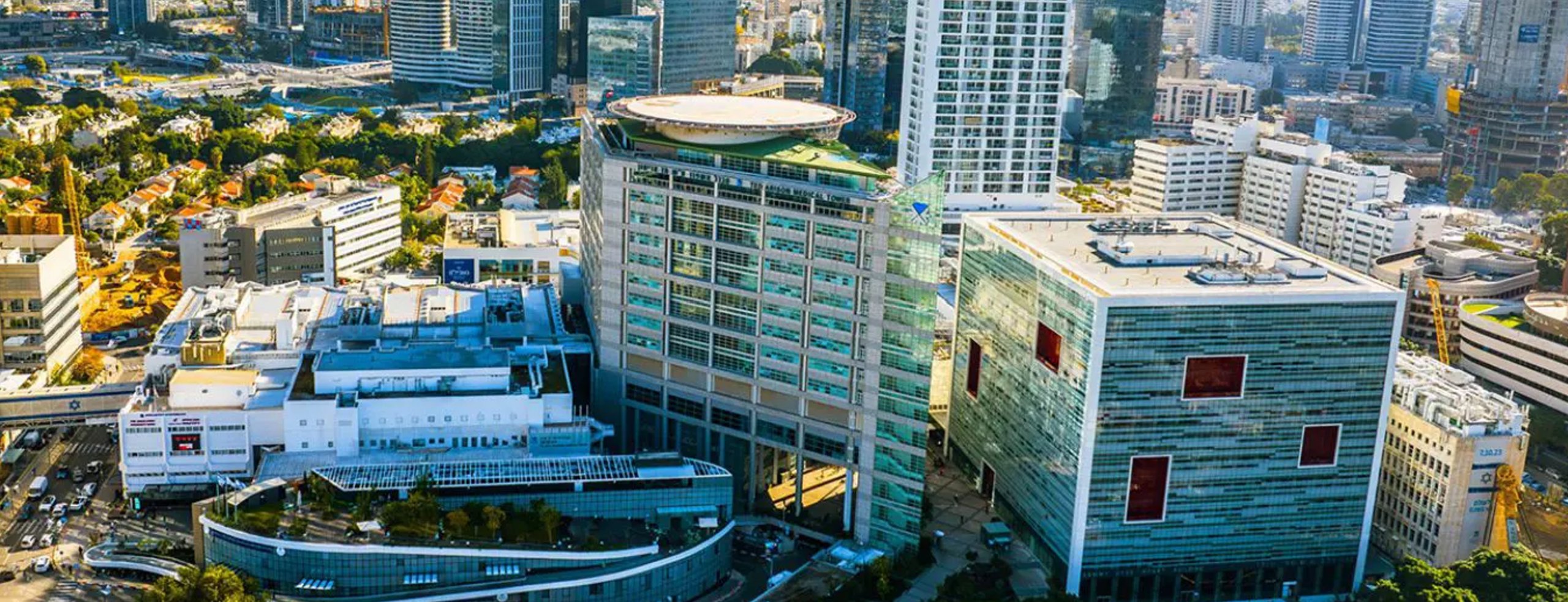
Sourasky Medical Center (TASMC) is a 1,500-bed governmental academic medical center affiliated with the Faculty of Medicine at Tel Aviv University. It serves approximately 400,000 patients per year and hosts 1.8 million patient visits annually. The hospital’s hematology division is one of the largest in the region, providing the full spectrum of clinical services - hemato-oncology, bone marrow transplantation, CAR-T cell therapy, and outpatient hematology clinics - supported by a multi-unit laboratory that includes routine hematology, flow cytometry, specialty coagulation, immunohematology, stem cell processing, and molecular biology.
The hematology laboratory processes approximately 1,300 complete blood counts per day. Around 70 peripheral blood smears are required daily - split roughly evenly between automated reflex flags and clinician-ordered reviews.
The challenge
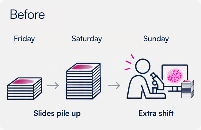
Like most hospital laboratories, Sourasky Medical Center operated without full peripheral blood smear review capacity over the weekend. At TASMC, the work week runs Sunday through Thursday, so Friday and Saturday are the weekend and Sunday is the first working day - equivalent to Monday in most countries. Only urgent stat samples - suspected leukemia, suspected thrombotic thrombocytopenic purpura - were processed on Friday and Saturday. Everything else accumulated. By the time the first working day of the week arrived, the lab faced a backlog requiring an additional eight-hour shift just to catch up.
The impact cascaded forward. Average turnaround time for Friday samples reached over 45 hours. The first-weekday backlog pushed average turnaround to nearly eight hours even after the extra shift began.
Beyond the weekend problem, the lab faced a limitation common to any site using traditional cell-snapshot digital analyzers: roughly 40% of cases still required reversion to the manual microscope - for platelet clump detection at the feathered edge, for red blood cell morphology assessment, for confirmation of flags. True end-to-end digital review wasn’t possible.
Weekend workflow bottleneck
- InstitutionSourasky Medical Center (TASMC)
- Type1,500-bed governmental academic medical center
- Patient volume~400,000 patients/year; 1.8 million visits
- Peripheral blood smears~70 per day
- StatusWorld's first Full-Field digital morphology installation

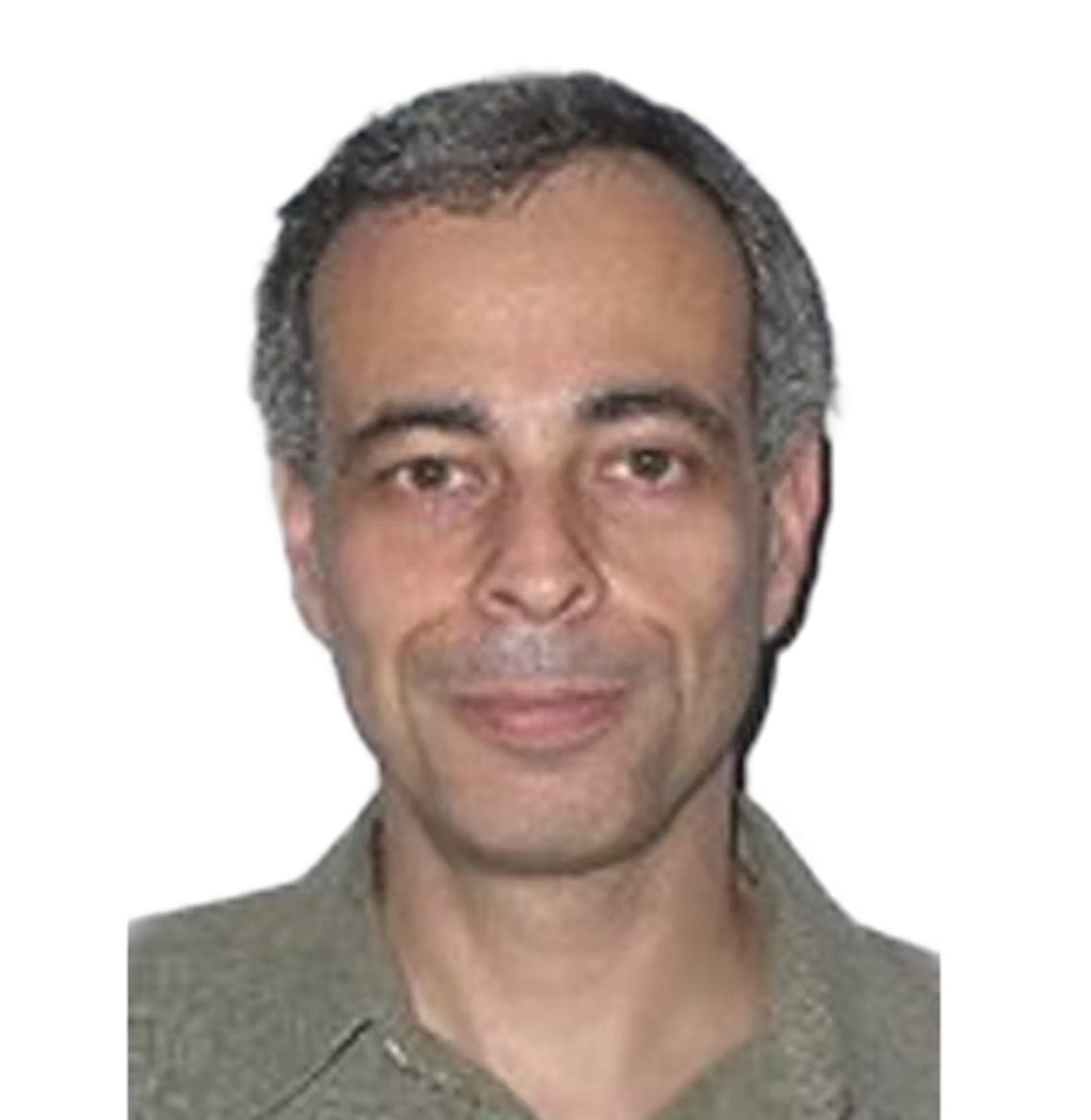
We broke the triangle of the slide, the microscope, and the morphologist. Previously, all three had to be on the same site together. Now they are separated - and that changed everything.
Dr. Ben-Zion KatzDirector of the Hematology Laboratory, Sourasky Medical Center
The solution
Dr. Ben-Zion Katz, Director of the Hematology Laboratory and a Senior Lecturer at Tel Aviv University’s Faculty of Medicine, led the deployment of Scopio’s Full-Field digital morphology platform* - the first such installation in a live clinical setting worldwide.
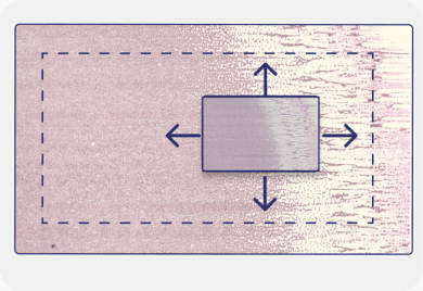
The key difference from existing digital systems wasn’t just image quality - it was coverage. Where cell-snapshot analyzers capture a narrow field of view and require manual fallback for edge-of-slide findings, Scopio’s approach scans all clinically relevant areas of the peripheral blood smear at 100x resolution, including the monolayer and feathered edge. Platelet clumps, rare cell forms, and morphological detail at the margins are no longer reasons to pick up the microscope.
That shift - from partial digital to fully digital - changed what was operationally possible. Once the review could happen entirely on screen, it no longer had to happen in the lab.
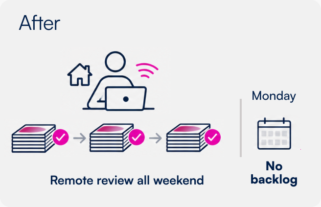
Scopio’s web-based platform meant that certified morphologists could review slides remotely*, from home, during weekends and outside regular hours. Dr. Katz restructured the weekend workflow: morphologists were enabled to perform the analysis remotely, compensated for extra hours, and the backlog was addressed at its source rather than downstream.
“It fully eliminates the need for additional manual microscopy.”
Dr. Ben-Zion Katz, Director of the Hematology Laboratory, Sourasky Medical Center
The results
The lab measured outcomes across two matched five-month periods - before and after implementing remote digital morphology review - covering more than 10,000 samples. The results were published in Applied Clinical Informatics (Katz et al., 2022).
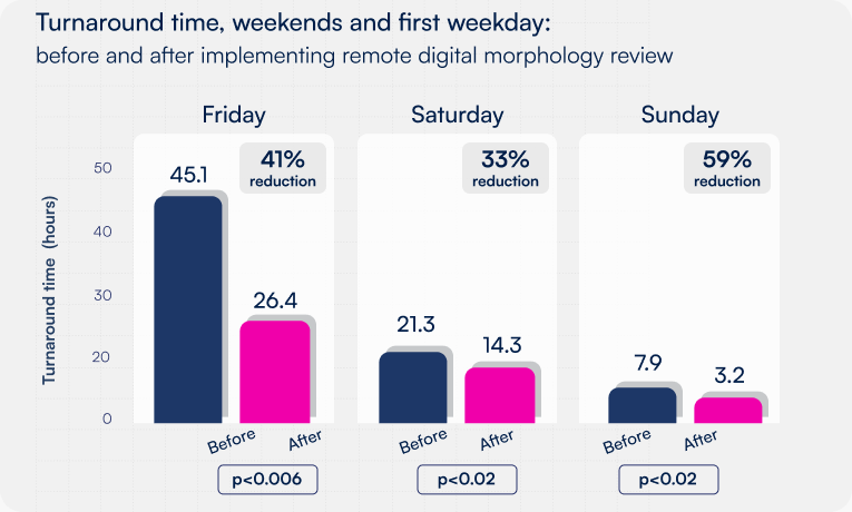
Turnaround time, weekends and first weekday:
- 41% reduction in average Friday turnaround time (from 45.1 hours to 26.4 hours; p<0.006)
- 33% reduction in average Saturday turnaround time (from 21.3 hours to 14.3 hours; p<0.02)
- 59% reduction in average Sunday turnaround time (from 7.9 hours to 3.2 hours; p<0.02)
Lab-wide impact:
- 15.8% reduction in overall morphology turnaround time across the laboratory - statistically significant, and achieved with no increase in overall workload
- 36 staff hours saved per month by eliminating the first-weekday morning extra shift
- ~12 net hours saved per month after accounting for the additional remote hours paid to weekend morphologists
The recurring extra shift - a persistent cost and scheduling burden - was eliminated entirely.
The workflow transformation went beyond the data. Every morphology report issued by the lab now includes a direct link to the digital slide, accessible from anywhere on the hospital network. Hematologists no longer travel to the lab to review a sample. Clinicians on call at night or over the weekend can view the slide from home and give a result without leaving.
When the COVID-19 pandemic forced the entire morphology team to isolate simultaneously, the lab continued operating without interruption. Staff reviewed samples remotely throughout the week.
The near-zero reversion rate to manual microscopy was another landmark outcome. Scopio’s platform covers everything the manual review covered - meaning the microscope is no longer a fallback.
“I simply could connect from my computer at my desk in the office and do it simultaneously. It’s unbelievable. It’s very easy.”
Dr. Ben-Zion Katz
Beyond workflow: a platform for research
The adoption of digital morphology at Sourasky Medical Center didn’t stop at operational improvement. The ability to capture thousands of cells per slide at 100x resolution - routinely, across every sample - opened research territory that simply wasn’t accessible with conventional digital or manual methods.
CAR-T cell monitoring in peripheral blood
Monitoring chimeric antigen receptor T-cell (CAR-T) therapy in patients with relapsed or refractory diffuse large B-cell lymphoma typically relies on PCR or flow cytometry - both expensive, variable across products, and requiring specialist protocols. Neither method provides information about the functionality of the cells being measured. Dr. Katz’s team used Scopio’s platform to explore whether morphological surveillance of peripheral blood smears could track CAR-T cell dynamics after infusion - offering a simpler, faster, and less costly alternative.
The challenge was sensitivity. In leukopenic patients receiving CAR-T therapy, the circulating white cell count drops sharply, leaving very few cells to analyze. In a head-to-head comparison across 77 samples from 18 patients, Scopio’s platform retrieved an average of 303 white blood cells per sample compared to 54.4 using a cell-snapshot system - a statistically significant difference (p < 0.001). For patients with very low white counts in the first days post-infusion, that difference determines whether meaningful analysis is possible at all.
Across 166 consecutive peripheral blood smears from 26 patients, the team identified five distinct CAR-T cell morphological subtypes in circulation - quiescent, activated, apoptotic, multinucleated, and mitotic. Total CAR-T cell counts on day 5 post-infusion were significantly higher in patients who achieved complete response at one month (17.1 vs. 2.8 cells per sample, p = 0.018), and sustained presence of the activated morphology subtype through days 1-14 was also associated with complete response. Activated morphology CAR-T cells at day 14 were significantly correlated with cytokine release syndrome duration.
The study - published in Cancers (Fridberg, Katz et al., 2023) following earlier presentation at the European Hematology Association Annual Congress - represents the first morphological characterization of CAR-T cells in peripheral blood, positioning digital peripheral blood smear review as a practical complement to existing molecular monitoring methods. Read the full study on Scopio’s Clinical Impact page.

The clinical context: 22-year-old male referred with suspected lymphoma and paravertebral lymphadenopathy. CBC unremarkable except mild leukocytosis.
Quantitative morphometry in myelodysplastic syndrome
Current diagnosis of myelodysplastic syndrome (MDS) depends on invasive bone marrow aspirate followed by subjective morphological assessment. Dr. Katz’s team applied Scopio’s platform to peripheral blood smear analysis in MDS - asking whether quantitative, large-scale morphometric measurement could complement or reduce reliance on manual grading.
The study analyzed 32 MDS and 30 age-matched control samples, evaluating three morphological features with known significance in MDS: blast percentage (per 100 or 1,000 white blood cells), neutrophil cytoplasmic granulation (per 1,000 neutrophils), and red blood cell morphology (at least 150,000 red blood cells per sample).
Key findings: when blasts were counted per 1,000 white blood cells rather than the conventional 100, detection rose from 41% to 84% of MDS samples - a meaningful sensitivity gain enabled only by the platform’s high cell yield. Neutrophil granulation, assessed via a quantitative Granulation Index, showed statistically significant differences between MDS and control samples (p<0.0001), with two distinct sub-populations identified in some MDS samples - a potential fingerprint of coexisting normal and abnormal clones. Red blood cell size and deformation were significantly elevated in MDS versus controls (p<0.000001).
This work - toward what Dr. Katz describes as a shift from morphology to morphometry - points to a future where peripheral blood reports include numerical parameters with reference ranges, rather than semi-quantitative grading. Read the full poster.
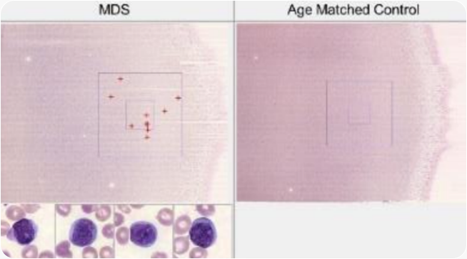
The Granulation Index histograms from the MDS poster- normal distribution vs. MDS bimodal distribution, side by side
Looking ahead
Dr. Katz’s vision for the field extends the current platform into what he calls morphometry: moving from descriptive grading of cellular features (e.g., reporting toxic granulation as mild, moderate, or severe) to precise numerical parameters - granulation index, deformation percentage, macrocyte fraction - reported with reference ranges alongside the standard differential count.
In an MDS report built on Scopio’s platform, that means a clinician sees not just “hypogranulation present” but a quantified granulation index plotted against a normal distribution, with a flagged sub-population of abnormal neutrophils visible in a histogram. For blast enumeration, it means sensitivity measured at 1,000 cells rather than 100 - a tenfold increase that changes what the peripheral blood smear can detect.
This trajectory aligns with the broader evolution underway at Scopio. Complete Blood Morphology (CBM)™ - currently in multi-center validation - aims to fully automate routine peripheral blood smear analysis, delivering standardized quantitative reports directly to the laboratory information system and eliminating routine morphology review entirely. Dr. Katz has described CBM as potentially “just the tip of the iceberg, because digital morphology using the Full-Field technology can provide new horizons for the development of new digital applications in the field of morphology.”
Watch Dr. Katz present the morphometry concept in the From Morphology to Morphometry webinar.
“This is just the tip of the iceberg. Once we have the Full-Field in our hands, the numerical side of applications is very large.”
Dr. Ben-Zion Katz
The evolution

Manual Microscopy: Samples reviewed only in the lab

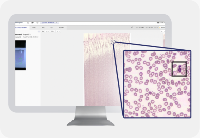
Digital Cell Morphology:
Samples reviewed and signed off from anywhere
Autonomous Cell Morphology and Reporting: AI-powered Analysis of 10x more cells
References
- Katz BZ, Benisty D, Sayegh Y, Lamm I, Avivi I. Remote digital microscopy improves hematology laboratory workflow by reducing peripheral blood smear analysis turnaround time. Applied Clinical Informatics. 2022 Oct 8. Scopio clinical impact page | PubMed Central full text
- Fridberg G, Horn G, Globerson Levin A, Benisty D, Kay S, Glait-Santar C, Perry C, Ram R, Avivi I, Katz B-Z. The clinical significance of circulating lymphocytes morphology in diffuse large B-cell lymphoma as determined by a novel, highly sensitive microscopy. Cancers. 2023;15(23):5611. Scopio clinical impact page | MDPI full text
- Katz B-Z, Karny S, Shimoni H, et al. Automated digital morphometry of peripheral blood smears detects both infrequent events and cellular population patterns in myelodysplastic syndrome. Scopio clinical impact page
Related resources
- Watch: From Morphology to Morphometry - ISLH 2024 Workshop - Dr. Ben-Zion Katz
- Watch: ISLH 2021 - Evaluation of the Scopio X100 Full-Field Peripheral Blood Smear Platform - Dr. Ben-Zion Katz
- Full-Field peripheral blood smear review
- Complete Blood Morphology (CBM) - autonomous analysis
- Browse all clinical evidence
More stories




- Scopio Labs’ Full-Field Peripheral Blood Smear application is CE marked and FDA-cleared, and it’s commercially available across the U.S., UK and Europe and other territories.
- Scopio’s Full-Field remote capabilities are available through the secure hospital network.