
- Customer Stories
- Hematopathology Hamburg
How Hematopathology Hamburg Brought AI-Assisted Digital Morphology into Routine Leukemia and Lymphoma Diagnostics
A specialized German hematopathology institute integrates Full-Field digital imaging for peripheral blood and bone marrow aspirate review - streamlining workflows, reducing manual handling, and giving experts the freedom to work from anywhere.

cases processed
per year
annual case
volume growth
from staining
to sign-off
location-independent
expert review
About Hematopathology Hamburg
Hematopathology Hamburg (HpH) is a private medical laboratory specializing in leukemia and lymphoma diagnostics. The institute handles approximately 40,000 cases annually, with case volumes growing by an average of 5% year over year.
As a specialized reference lab, HpH receives a higher proportion of pathological cases than a typical clinical laboratory. Nearly every case requires morphology assessment, and most require combined diagnostic procedures - morphology, flow cytometry, molecular analysis, and histology - synthesized into a single integrated report.
- InstitutionHematopathology Hamburg (HpH)
- LocationHamburg, Germany
- TypePrivate specialty reference laboratory
- SpecializationLeukemia and lymphoma diagnostics
- Annual volume~40,000 cases per year
The challenge
HpH faced a combination of pressures familiar to specialized hematopathology labs across Europe:
Growing workload and rising case complexity
Case volumes were increasing year over year, with individual cases requiring multi-step diagnostic algorithms and synthesis of results from multiple modalities. Throughput needed to keep pace without compromising quality.
Excessive manual handling in the lab
A thorough workflow analysis revealed dozens of manual steps in routine case processing - including manual matching of paper records to corresponding samples and physical transport of records and samples between departments. Many of these movements within the lab were redundant and added error risk.
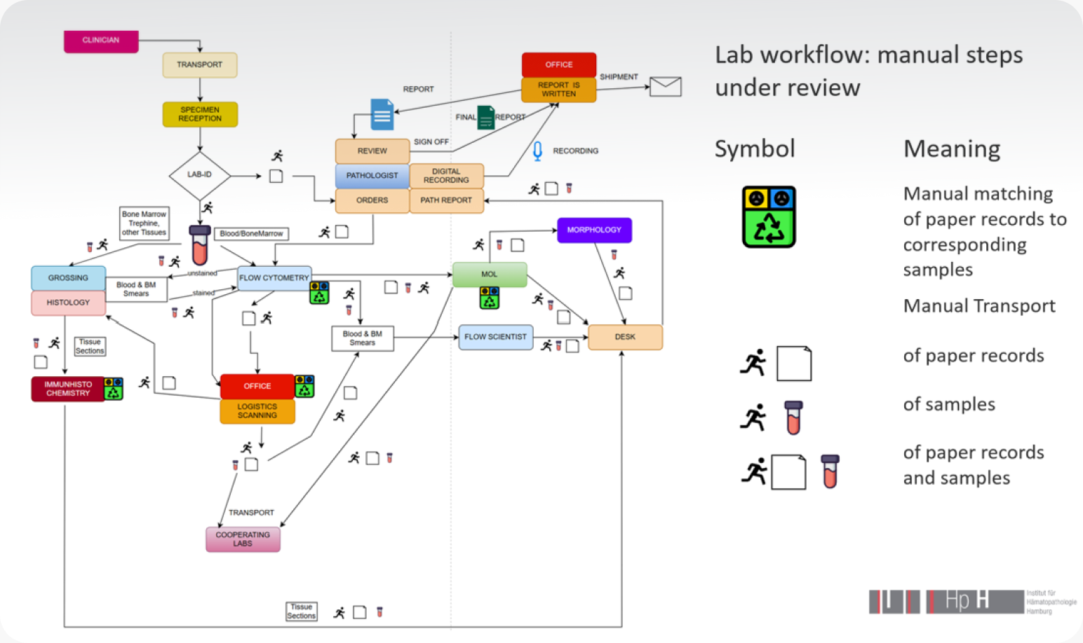
“We had so many manual steps. The papers had to be sorted to glass slides, the slides had to be brought to another facility to be stained, then brought back. After that, everything had to be collected in one place and physically brought next to the microscope. That's miles of movement every day, and a lot of time.”
Dr. Holger Hauspurg, Head of Hematopathology,
Hematopathology Hamburg
Demographic pressure on specialist availability
Like many European hematopathology labs, HpH faced the dual reality of an aging patient population driving more cases and fewer trained specialists available to review them.
Demand for location flexibility
Highly trained morphologists were tied to on-site microscopes, limiting flexibility for evening, weekend, and urgent case coverage. Location independence had become a priority for staff retention.
Unsatisfactory results from other digital scanners
Before finding Scopio, the team had evaluated other digital scanning devices but found them insufficient for both peripheral blood and bone marrow aspirate smears.


We were happy to find Scopio because all other scanning devices we tested for peripheral blood and especially for bone marrow aspirate smears had underwhelming results.
Dr. Holger HauspurgHead of Hematopathology, Hematopathology Hamburg
The solution
HpH took a two-step approach to digital transformation.
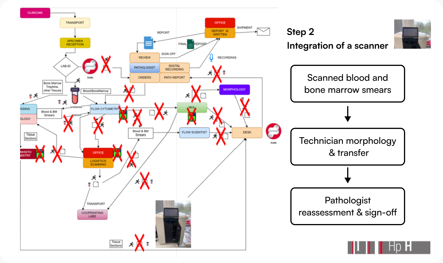
First, the team built its own middleware platform - called Floki - to centralize all paperwork associated with each sample and digitize record matching across departments. This single change eliminated a major source of manual effort and significantly improved connectivity between flow cytometry, molecular pathology, and other lab sections, while enabling end-to-end sample tracking.
Then, the team integrated Scopio’s Full-Field digital imaging platform for peripheral blood and bone marrow aspirate morphology. With Scopio, the morphology assessment process was streamlined into three steps following slide staining:
- Scan the peripheral blood or bone marrow aspirate smear at 100x resolution
- Technician morphology review - verify the AI pre-classification and reclassify cells as needed
- Pathologist reassessment and sign-off - results transfer automatically via HL7 interface to the Floki middleware, accessible in one click
“Once a case is signed off, a message with the result of the differential is transferred via an HL7 interface to our Floki middleware. From the middleware, you can access it via one click and land straightforward in the whole slide imaging review.”
Dr. Holger Hauspurg, Head of Hematopathology,
Hematopathology Hamburg
Results
Streamlined, digitally connected processes
The integrated Floki-plus-Scopio workflow replaced dozens of manual handling steps with a connected digital pipeline. The workflow reduces reliance on manual paper-to-slide matching, and physical transport routes between lab sections have been substantially reduced.
Reduced workload through AI-assisted morphology
The AI pre-classification of cells provides a strong starting point for differential review - particularly valuable when a hypocellular smear makes finding 100 cells for a manual differential difficult.
True location independence for expert reviewers
Perhaps the most transformative result has been the freedom for HpH’s specialized morphologists to work from anywhere. Cases no longer require physical presence at the microscope. Urgent reviews can happen in real time, regardless of where the expert is located.
The most memorable example: on a Saturday morning, Dr. Hauspurg received a call about a suspected acute leukemia case while at the supermarket. He stepped aside to a quiet spot and confirmed the diagnosis of acute promyelocytic leukemia on his phone - in under a minute. Without the digital workflow, the same diagnosis would have required a drive to the lab and more than an hour at the microscope.
The team confirmed acute promyelocytic leukemia via flow cytometry and molecular analysis the same day - significantly accelerating the path to treatment for a clinical emergency.
Confidence in whole slide imaging for bone marrow aspirate
For bone marrow aspirate cases, Scopio’s Full-Field whole slide imaging has proven essential - particularly for catching findings that AI-selected differential areas can miss.
In one recent case, a 22-year-old male was referred with suspected lymphoma. The AI’s pre-selected differentiation areas didn’t capture the cluster of malignant cells, but Dr. Hauspurg identified them on the whole slide review. The diagnosis turned out to be a rare embryonal neuroectodermal tumor of the testis with bone marrow metastases - a finding that could have been missed without comprehensive slide coverage.
Reliable identification of difficult stains
The platform handles Prussian blue staining - historically one of the most difficult stains for scanning devices - with the clarity needed for myelodysplastic neoplasm diagnosis. In a recent case of MDS with ring sideroblasts, the team identified ring sideroblasts with ease using the Prussian blue scan, alongside merged Romanovsky-stained slides under a single patient ID.
“It's very appreciated to have an AI-assisted differentiation up front that you can use as a starting point for your refinement.”
Dr. Holger Hauspurg, Head of Hematopathology,
Hematopathology Hamburg
“Without the digital workflow, I would have had to go to the lab and reassess the diagnosis on my microscope, and it would have taken me more than one hour.”
Dr. Holger Hauspurg, Head of Hematopathology,
Hematopathology Hamburg
“In my opinion, single-cell analysis alone isn't reliable for a bone marrow aspirate smear. You're seeing one branch of one tree – you miss the whole picture.”
Dr. Holger Hauspurg, Head of Hematopathology,
Hematopathology Hamburg
Clinical cases
Acute promyelocytic leukemia diagnosed remotely* on a Saturday morning
A 36-year-old male presented with leukocytosis, blasts in peripheral blood, and thrombocytopenia. Dr. Hauspurg reviewed the case remotely on his phone from a supermarket parking lot, confirming acute promyelocytic leukemia in under a minute. The diagnosis was later confirmed by flow cytometry and PML::RARA molecular analysis - with treatment initiated dramatically faster than would have been possible with a microscope-based workflow.
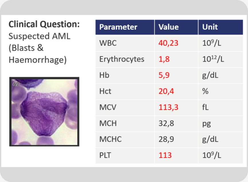
The clinical context: 36-year-old male, WBC 40.23 × 10⁹/L, blasts in peripheral blood, anemia, thrombocytopenia.

Full-Field view of the APL slide.
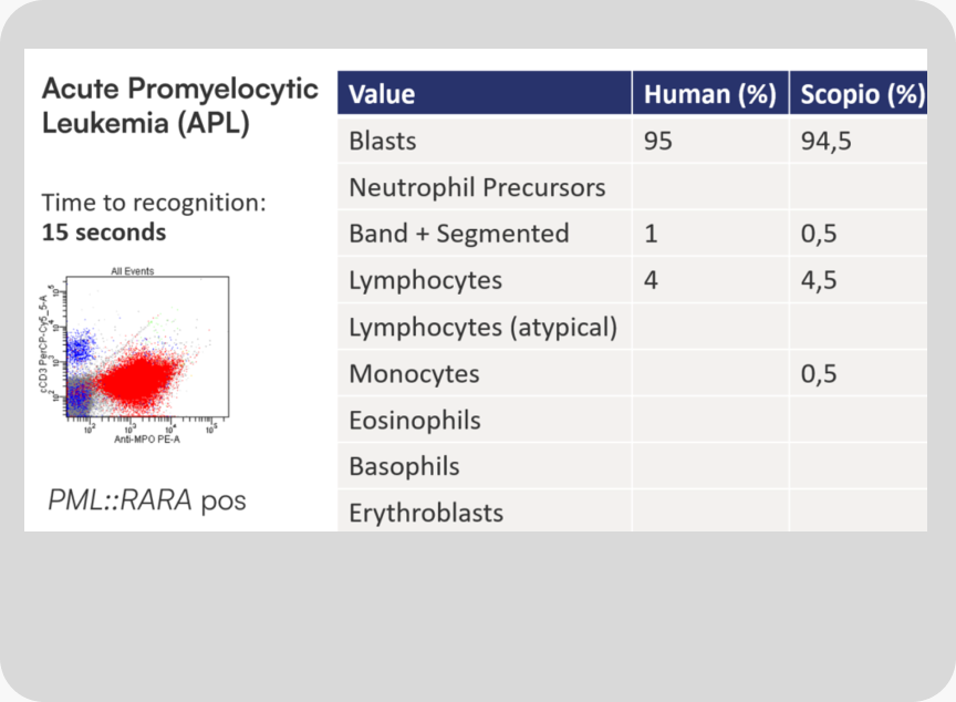
Diagnosis confirmed: acute promyelocytic leukemia, PML::RARA positive. Differential count completed in 4 min 22 sec.
Bone marrow metastases from a rare embryonal neuroectodermal tumor
A 22-year-old male was referred with suspected lymphoma and paravertebral lymphadenopathy. The AI’s pre-selected differential areas missed the area of clinical interest - but on whole slide review, Dr. Hauspurg identified a cluster of malignant cells that turned out to be metastases from an embryonal neuroectodermal tumor of the testis. The case became a teaching example of why whole slide review remains essential for bone marrow aspirate.
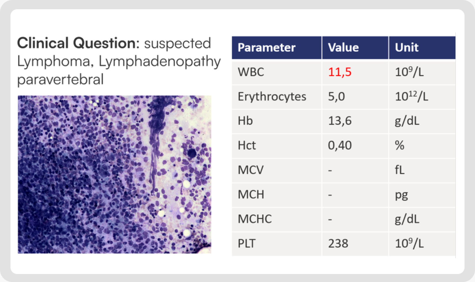
The clinical context: 22-year-old male referred with suspected lymphoma and paravertebral lymphadenopathy. CBC unremarkable except mild leukocytosis.
Looking ahead
HpH is actively expanding its integration of Scopio’s platform. Two priorities are on the immediate roadmap:
Deep integration of bone marrow aspirate morphology into Floki
The team plans to extend the same middleware integration that exists for peripheral blood - transferring myelogram differentials directly into HpH’s pathology reports.
Connectivity between Scopio and HpH’s digital pathology platform
Because many cases combine peripheral blood smears, bone marrow aspirate smears, and histology slides, the team is working to bring all morphology into a single integrated workflow rather than working across multiple platforms.
“We have a lot of cases that are combined peripheral blood smears, bone marrow aspirate smears, and histology slides, and it is not a good idea to work on one case with two or even more different platforms.”
- Dr. Holger Hauspurg, Head of Hematopathology, Hematopathology Hamburg
The human-in-the-loop principle remains central to how HpH thinks about AI in diagnostics.
“With the AI morphology you have a good starting point, but the human in the loop is still needed.”
- Dr. Holger Hauspurg, Head of Hematopathology, Hematopathology Hamburg
The clinical context: 22-year-old male referred with suspected lymphoma and paravertebral lymphadenopathy. CBC unremarkable except mild leukocytosis.
From diagnosing acute promyelocytic leukemia from a supermarket parking lot to catching rare bone marrow metastases on whole slide review, Dr. Holger Hauspurg shares how Hematopathology Hamburg integrated AI-assisted digital morphology into routine leukemia and lymphoma diagnostics. Watch the full ISLH 2026 workshop.
“Digital imaging systems will increasingly change the diagnostic environment. With the implementation of a digital workflow and AI-supported blood morphology, we have streamlined our processes, reduced the probability of errors, reduced the workload of our morphologists, and reached independence from location for our highly trained staff - leading to higher satisfaction with their work environment.”
Dr. Holger Hauspurg, Head of Hematopathology,
Hematopathology Hamburg
More stories




- *Scopio’s Full-Field Bone Marrow Aspirate Application is CE-marked and cleared for sale in CE countries as well as in additional regions. Pending FDA clearance in the US.
- *Scopio’s Full-Field Peripheral Blood Smear Application is FDA-cleared and CE-marked.
- *Scopio’s Full-Field remote capabilities are available through the secure hospital network.