

Wherever, whenever, review and
report from your browser
Reduce the manual burden and standardize
bone marrow review across every shift and site
CE-IVDR certified. Pending FDA clearance in the US.
*Pabon et al., Beckman Coulter, 2026.
The Full-Field difference
A fully digital review
No slide transport.
No microscope dependencies.
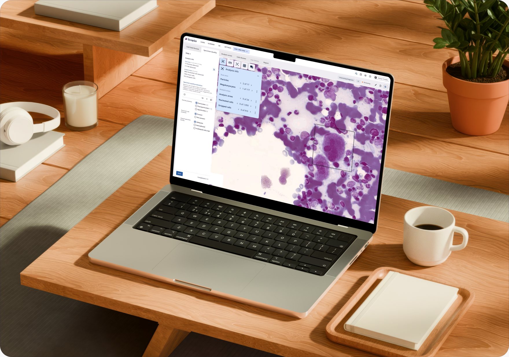
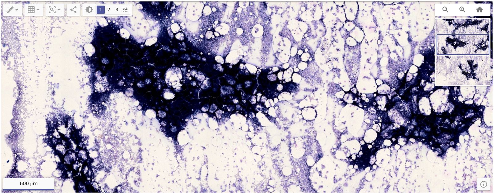
Remote team members access the entire digital sample and toolset from any workstation*, enabling consultations and more flexible staffing.
*through the secure hospital network.
Already in routine clinical use
Real-world adoption and peer-reviewed validation
Real-world adoption

4,000+
bone marrow cases
reviewed with Full-Field
at Mount Sinai Health System.
In routine clinical use since September 2024, following internal validation on a 60-case cohort including MDS and AML cases.
Independent validation
Validated in Modern Pathology.
Peer-reviewed multicenter study across 795 bone marrow aspirate specimens. Bagg et al., 2024.
Read the studyBringing on Scopio completed the digitization of our hematology lab, clearing the bone marrow workflow that has historically been a major manual bottleneck.

I have been waiting for such a device for almost five years.

Remote access to high-resolution web-based images is an important feature: when we work from home, when we are on call on weekends.



Frequently asked questions
What labs ask when evaluating Full-Field

The Scopio Full-Field Bone Marrow Aspirate™ Application is an end-to-end digital workflow for bone marrow review. It uses Full-Field imaging to capture the entire slide at 100x, AI to pre-classify nucleated cells into 17 categories, and a browser-based interface so experts can review and sign off regardless of their location.
The Scopio Full-Field Bone Marrow Aspirate Application is CE-IVDR Class C certified for the European Union and additional regions. Pending FDA clearance in the US.
Yes. The Full-Field BMA Application supports bi-directional HL7 connectivity with your laboratory information system. Bone marrow scans export natively in DICOM to any compatible PACS or image management system.
No. AI pre-classifies cells; the hematopathologist verifies findings and signs off on every case. The expert maintains absolute diagnostic authority.
The Scopio Full-Field BMA Application supports smear/film, imprint/touch, and squash/crush preparations. It works with Romanowsky stains (Wright-Giemsa, May-Grünwald Giemsa, and others) and with Prussian Blue for iron stores evaluation.
Yes. The Scopio platform is browser-based, so experts can review and sign off on bone marrow cases from any workstation.*
Reviewers tag cells as abnormal during review. The system calculates the percentage of tagged cells within each cell class, supporting dysplasia assessment.
AI pre-classification provides a standardized starting point for every differential, applied consistently across reviewers, sites, and shifts. The expert verifies and confirms findings, maintaining final diagnostic authority.
Full-Field is the only platform that captures the entire bone marrow slide at 100x. Reviewers navigate the sample as they would on a microscope, but entirely on screen, completing the full workflow from slide to sign-off. Book a meeting to see how it compares to your current workflow.