
- Customer Stories
- Bone Marrow Aspirate
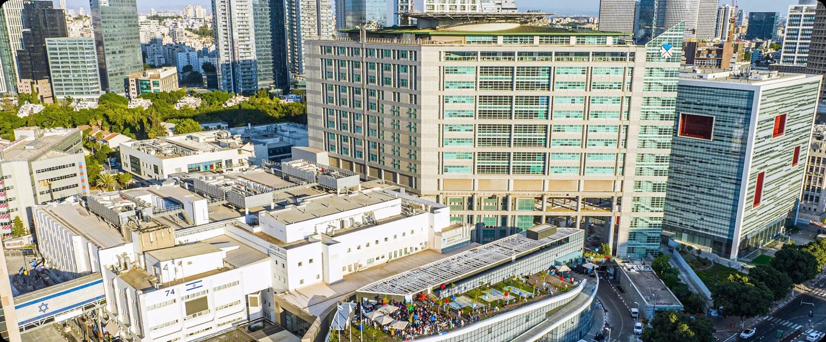
Two Minutes to Sign-Off: How Sourasky Medical Center Cut Bone Marrow Aspirate Review Time by Up to 70%
Faster sign-off, less time counting cells, and urgent cases reviewed the same evening: how Scopio’s Full-Field Bone Marrow Aspirate Application changed the workflow at Sourasky.

Less review time for normal and acute leukemia cases
Agreement on blast identification in acute leukemia
For urgent cases
About Sourasky Medical Center
Sourasky Medical Center (TASMC) is a 1,500-bed governmental academic medical center affiliated with the Faculty of Medicine at Tel Aviv University. Its hematology division covers the full spectrum of care - hemato-oncology, bone marrow transplantation, CAR-T cell therapy and outpatient clinics - which makes bone marrow evaluation a daily, high-stakes part of the workload.
Sourasky has a long history with digital morphology. Its hematology laboratory was the world’s first site to deploy Scopio’s Full-Field peripheral blood smear platform (read that story), and its surgical pathology service has worked on digital scanners since 2017.
In 2025, Sourasky’s hematopathology team brought bone marrow aspirate review onto the same platform, adding Scopio’s Full-Field Bone Marrow Aspirate Application.
The challenge
Bone marrow aspirate review was the last part of Sourasky’s hematopathology work still done entirely by hand.
Time spent counting, not interpreting
Even a normal marrow required a full manual differential: searching for fields, choosing representative areas and counting cells, before any interpretation began. Across 80-100 cases a week, that time added up.
“Before Scopio, just counting 100 cells took five to seven minutes. You search for a field, you look, you choose the representative areas… it takes time.” Sara Feldstein, M.D., Hematopathologist and Researcher, Sourasky Medical Center
Tied to the lab
Because the glass couldn’t leave the lab, neither could the hematopathologist. Every case meant sitting at the bench in the lab, and urgent samples that arrived in the evening waited until the next morning.
A differential limited by time and endurance
A manual count gives an impression from a handful of fields and 100 cells - as many as one person can reasonably count. The ratio is only as good as that sample.
Rare diseases, hard to triage
For ultra-rare entities, the flow cytometry team needs morphology to know which panel to run. Without an early look at the smear, the case waited until morning, or markers and material risked being spent on the wrong panel.
- InstitutionSourasky Medical Center (TASMC)
- LocationTel Aviv
- Type1,500-bed governmental academic medical center
- ServiceHematopathology
- Volume~80-100 bone marrow aspirates per week
- Live since2025
The solution
The hematology laboratory scans two aspirate smears per case on the Scopio X100HT. The hematopathologist opens the case in a web-based viewer, marks the areas to analyze on each slide, reviews the AI-supported differential alongside the full-resolution image, and combines both slides into a single assessment.
AI pre-classification, expert sign-off
Scopio’s AI detects and pre-classifies cells, groups them by lineage and calculates the myeloid-to-erythroid ratio. The hematopathologist verifies, reclassifies where needed, and signs off. Gross abnormalities stand out at a glance.
“What I really rely on is that it calculates the M:E ratio for me and gives me a rough division between the cell groups. If it suddenly sees eosinophilia, basophilia or thrombocytosis - if something gross stands out - it will catch it.” Sara Feldstein, M.D.
Many more cells per differential
Because the count isn’t limited by time or fatigue, the differential can be based on far more cells than a manual count of 100.
“I get much more information and the actual cell counts. I’m not limited by my time or endurance. Instead of counting 100 cells, it can count 5,000 cells for me and give me more information and a more accurate ratio.” Sara Feldstein, M.D.
One shared image for the whole team
Treating hematologists, hematopathologists and senior consultants all review the same digital case - from their own desks, or together on screen at the weekly consensus meeting.
“We had been waiting for this for a long time. It cuts down on commuting and hassle, and it really helps - there are areas that are harder for me to assess morphologically, and it actually classifies them beautifully.”
Sara Feldstein, M.D.


I can sign off on leukemia cases without coming in, in a minute to three.
Sara Feldstein, M.D.Hematopathologist and Researcher, Sourasky Medical Center
The results
Normal marrows signed off in two minutes
For a normal marrow, the hematopathologist glances at the case, checks that Scopio’s distribution matches what she sees, and signs off.
Acute leukemia signed off in one to three minutes
In overt AML and ALL, Dr. Feldstein matches the flow cytometry result to the digital differential and signs off without coming into the lab.
“In leukemia cases it’s also amazing - everything related to identifying blasts. If it’s frank blasts, like AML or ALL, it identifies them perfectly, and there is almost 100% correlation with what I identify. I can sign off on these cases without coming in, in a minute, two, three.” Sara Feldstein, M.D.
Urgent cases reviewed the same evening
When an urgent aspirate arrives after hours, the hematopathologist logs in remotely and gives a preliminary read that same evening.
“Many times, when an urgent test comes in during the evening, I connect from home, take a look and say, ‘Okay, this is what it looks like.’ Before, it would have waited until the morning.” Sara Feldstein, M.D.
Faster triage in rare disease
In ultra-rare entities, an early digital look at the smear guides the flow cytometry workup before markers and material are spent.
“In rare and ultra-rare diseases, like the case of BPDCN we had, if there’s a smear where nobody knows which direction to approach it from, Scopio helps immensely. Before, the case would have waited until the morning. Now that doesn’t happen.” Sara Feldstein, M.D.
Collaboration grounded in the findings
Treating hematologists often review the case in Scopio before the hematopathologist does, and the discussion starts from the same image.
“Why do I love pathology? Because there is proof. I have the slide - here, please take a look. If you want to argue, argue on the actual findings.” Sara Feldstein, M.D.
“If a case is a 'nothing burger', I can sign it off with Scopio in two minutes. I barely spend any time on counting. If it's a normal case, I trust it, and it's very fast.”
Sara Feldstein, M.D.
Clinical case: When morphology carries the diagnosis
Chronic myelomonocytic leukemia with a dendritic cell differential
The clinical context
A man in his early 40s was referred to hematology with progressive pancytopenia over six months and a relative monocytosis on his blood count.
What the aspirate showed
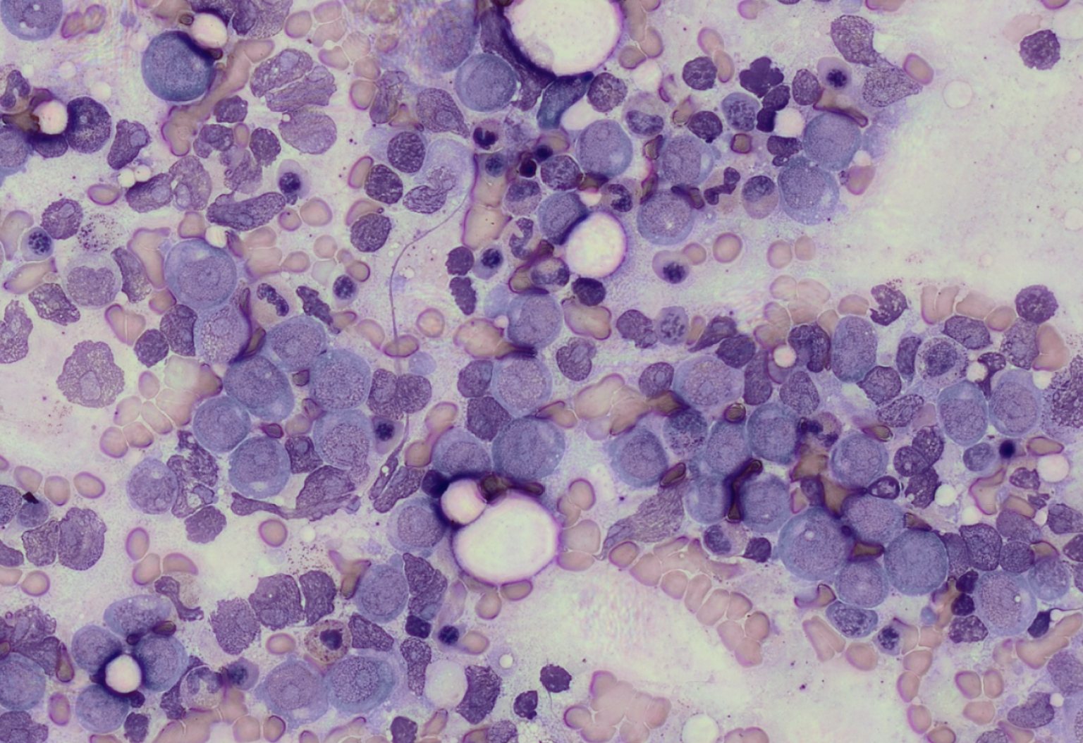
The bone marrow aspirate was scanned and reviewed in Scopio. It showed dysplastic trilineage hematopoiesis. The differential counted 4% myeloid blasts and 10% promonocytes. Flow cytometry, by contrast, detected only 2% myeloid blasts.
That gap is exactly where morphology matters. Promonocytes count as blast equivalents in chronic myelomonocytic leukemia (CMML), but flow cytometry cannot reliably separate them from mature monocytes. Only the aspirate differential captures them - and that count determines the subtype.
Putting the picture together
The biopsy was hypercellular (60-70%), with a 5:1 M:E ratio, dysplastic myeloid maturation, hypolobated megakaryocytes and clusters of monocytes. NGS detected IDH2 and SRSF2 mutations (both at roughly 40% VAF), and cytogenetics were normal. The case was presented at the weekly hematopathology consensus meeting, and the findings were reported as CMML, myelodysplastic subtype (CMML-1, WHO 5th edition).
When the differential widened
Weeks later, the patient developed widespread skin lesions. The skin biopsy showed monocytoid cells positive for CD4, CD123, CD56 (low), CD33 and lysozyme - raising the question of skin involvement by CMML versus a plasmacytoid dendritic cell proliferation, a close relative of BPDCN. Plasmacytoid dendritic cell markers were negative in the marrow by flow cytometry, and the case was sent for external expert consultation.
Where digital review made the difference
In a case like this, the flow cytometry team can’t know which panel to run until someone has looked at the cells. With the aspirate already in Scopio, the hematopathologist could review the morphology early and point the workup in the right direction, instead of the case waiting until the morning.
“With Scopio, I can look at the smear early, before the flow cytometry team spends markers and material on panels we don’t need.”
Sara Feldstein, M.D.
Looking ahead
Sourasky’s hematopathology team sees digital aspirate review as one step toward a fully connected workflow. Next on the list: sending the Scopio report directly into the laboratory information system, as surgical pathology already does, and building a digital teaching archive of starred cases for training and consultation.
More stories
 PERIPHERAL BLOOD SMEAR
How a 3-site NHS lab network went fully digital - and never looked back

 PERIPHERAL BLOOD SMEAR
Hematopathology was the last frontier for digital pathology. Here's how PathGroup got there
 PERIPHERAL BLOOD SMEAR
How a Swiss lab network cut peripheral blood smear review time by 57%
- *Scopio’s Full-Field Bone Marrow Aspirate Application is CE-marked and cleared for sale in CE countries as well as in additional regions. Pending FDA clearance in the US.
- *Scopio’s Full-Field Peripheral Blood Smear Application is FDA-cleared and CE-marked.
- *Scopio’s Full-Field remote capabilities are available through the secure hospital network.