
- Customer Stories
- The Center for Laboratory Medicine (ZLM)

How a Swiss Lab Network Cut Peripheral Blood Smear Review Time by 57% - and Standardized Morphology Across Six Sites
The Center for Laboratory Medicine (ZLM) - a 24-hour Swiss lab network serving hospitals, physicians, and university medical centers - deployed Full-Field digital morphology to eliminate manual microscopy bottlenecks, enable fully remote review*, and bring consistent quality to every site in its network.

Faster peripheral blood smear review turnaround at the central lab
Faster turnaround for satellite lab
cases reviewed remotely
Reduction in staff required at
the differential workstation
Connected through a single
digital workflow
About ZLM
The Center for Laboratory Medicine (Zentrum für Labormedizin, ZLM) provides 24-hour medical laboratory services to public and private hospitals, independent physicians, and university hospitals across Switzerland. ZLM’s main laboratory is located at the Cantonal Hospital in St. Gallen, with five additional satellite locations. Its hematology department specializes in morphology, flow cytometry, hemostasis, and immunohematology.
The network processes approximately 600 to 800 complete blood counts per day, with roughly 10% requiring morphology review - translating to 60 to 80 peripheral blood smear differentials daily across the system.
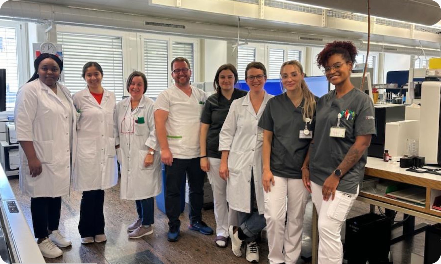
- InstitutionCenter for Laboratory Medicine (ZLM)
- LocationSt. Gallen, Switzerland
- TypeMulti-site reference laboratory network
- Network1 central hub lab + 5 satellite locations
- Pathologists600-800 CBCs per day; ~60-80 differentials
The challenge
ZLM faced rising demand for its services alongside a staffing shortage affecting the laboratory sector across Switzerland and beyond. Morphology analysis - already personnel- and expertise-intensive - had become a persistent bottleneck across the network.
Manual workflows slowing down the lab
Peripheral blood smear review using traditional light microscopy was labor-intensive and slow, requiring highly trained laboratory scientists to perform differentials and hematology experts to validate results. Two team members were dedicated to the differential workstation every day - a significant staffing commitment for a network under pressure.
Inconsistency across sites
Expertise varied across ZLM’s locations, particularly at the smaller satellite labs where staff saw difficult blood smears infrequently. Inter-variability among technicians was high, and the review process differed from site to site.
Physical slide transport adding hours to urgent cases
When a peripheral blood smear at a satellite lab required a secondary consultation, the physical slide had to be transported to the main lab in St. Gallen - a process that took one to 24 hours, or one to three hours for emergencies. During off-hours, this delay was even worse.
Remote reporting limited by cell-snapshot images*
Even for cases originating at the St. Gallen main lab, after-hours review was constrained. Lab staff would email a snapshot image of the slide to the expert on call. With only that partial view as a reference, consultants were confident enough to write an immediate remote report only about 50% of the time. The rest had to wait until the next day.

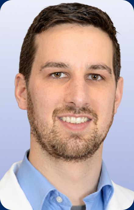
At the spoke sites, we have less specialized personnel. They see difficult blood smears infrequently. We wanted to be able to standardize the quality of patient care and bring morphology in every lab in our system to the same level.
Dr. Lukas GrafHead of Department of Clinical Chemistry, Hematology, Immunology, ZLM
The solution
Facing slow throughput and ongoing staffing pressures, ZLM leadership began evaluating digital morphology technologies. No existing solution fully addressed their needs until the team was introduced to Scopio and the concept of Full-Field digital morphology.
ZLM performed a rigorous validation on 50 peripheral blood smear samples, comparing manual microscopy against the Scopio X100 scanner with the Full-Field Peripheral Blood Smear Application*. The goal was to determine whether the system could partially or fully replace the manual microscope and provide a more efficient, consistent, and remotely accessible workflow for peripheral blood smear analysis.
The validation results confirmed that it could:
- High correlation for neutrophils, lymphocytes, monocytes, eosinophils, and platelets between Scopio and manual microscopy
- 99.77% agreement for red blood cell morphology between the test and reference method
- Platelet estimation accuracy: 94.89% efficiency, 90.00% sensitivity, 96.28% specificity
- White blood cell accuracy: 96.29% efficiency, 87.86% sensitivity, 97.62% specificity
- Overall end-user satisfaction rate of approximately 90%
ZLM has since deployed one X100HT scanner at the central lab in St. Gallen and three X100 scanners at satellite locations. Full-Field images can now be reviewed digitally from anywhere, immediately - giving reviewers the complete picture they need to report confidently without reverting to the microscope.
“Scopio was the only solution that fully met our needs”
“We looked for a digital solution that has remote access so that we are not limited to physical presence in the lab,” Richardson explains. “After successfully validating Scopio and implementing it in our main laboratory and different small spokes, it has really been life-changing in that we are able to standardize the quality of our differentials.”
“With Full-Field morphology, our experts and hematologists can perform analysis, then write and validate a report on a peripheral blood smear scan without reverting back to the manual microscope.”
Joyce Richardson, Lead Biomedical Scientist for Morphology and Immunohematology, Division of Hematology at ZLM
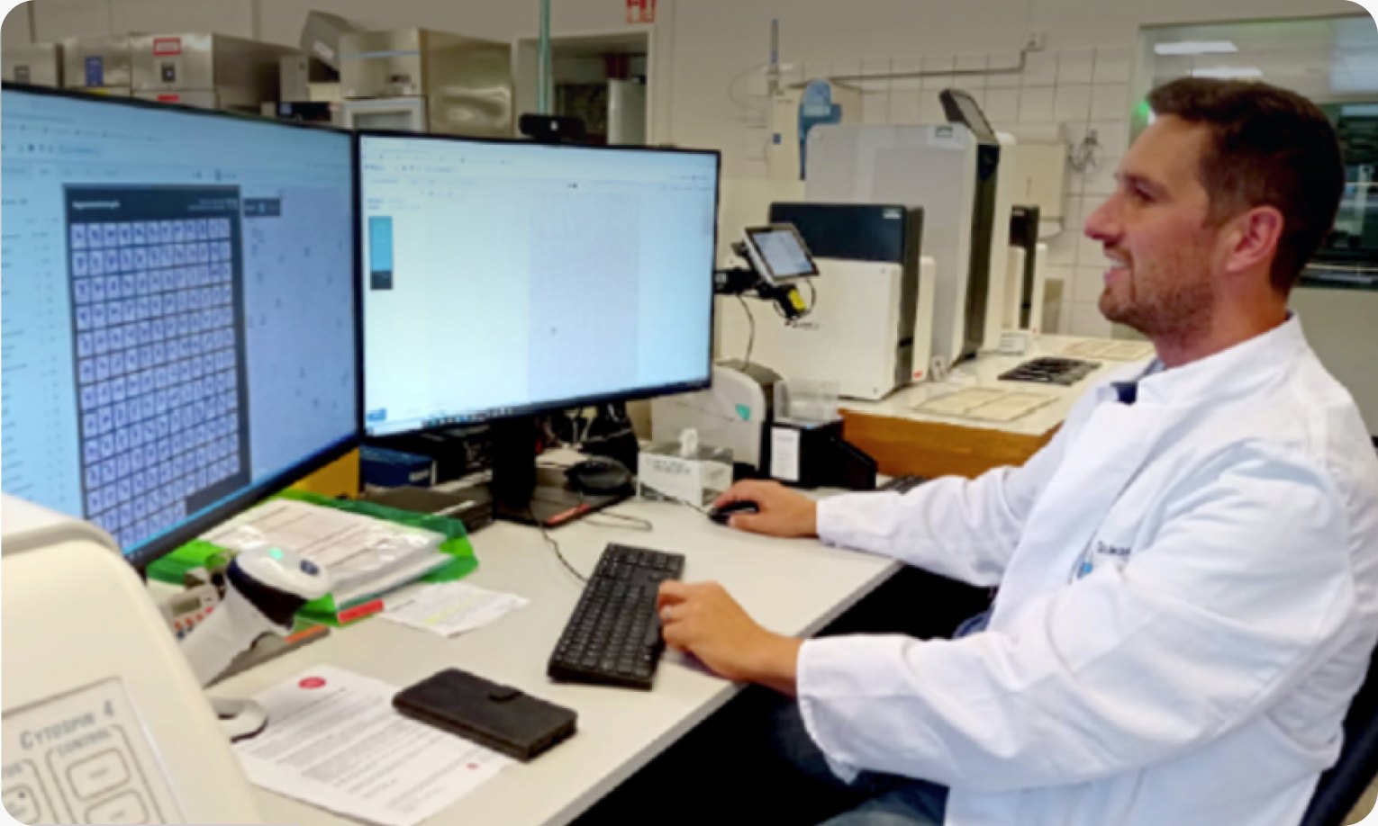
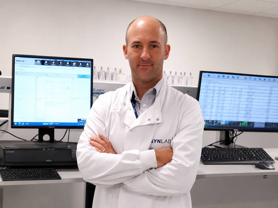
Dr. Lukas Graf in front of Scopio app
“The successful implementation of the Scopio system has enabled our hematologists to work remotely from anywhere around the world in real time. Being able to provide state-of-the-art morphology results 24/7 is a game changer for lab personnel, clinicians, and ultimately patients.”
Dr. Lukas Graf, Head of Department of Clinical Chemistry, Hematology, Immunology at ZLM
The Results
57% faster turnaround at the central lab
Full-Field digital morphology review at ZLM’s St. Gallen hub showed a 57% improvement in peripheral blood smear review turnaround time compared to manual light microscopy.
97% faster turnaround for satellite lab cases
For pathological samples originating at satellite laboratories and reviewed remotely at St. Gallen, turnaround time improved by 97%. Cases that previously required physical slide transport - adding hours or an entire day - are now reviewed within minutes of scanning.
50% reduction in staffing at the differential workstation
Before Scopio, ZLM required two team members dedicated to the differential workstation every day. After implementation, only one biomedical laboratory scientist is needed - freeing the equivalent of a full-time position for other work.
100% remote review capability
With the Full-Field image shared digitally, the consulting hematologist has everything they need to report confidently without returning to the lab. Where before only 50% of after-hours cases could be reported remotely from a snapshot, the rate is now 100%.
Standardized workflow across the network
The platform enabled ZLM to bring every lab in its system to the same standard. Satellite locations now have the same access to expert-level review as the central hub. Inter-variability across labs has decreased, and diagnoses at satellite sites are faster and more consistent.
“Specialized hematopathologists are based at the hub, so they don’t have to be present at small spokes to validate pathological samples,” says Richardson. “Having the Full-Field advantage allows us to have the confidence that a diagnosis based on the morphology you see on the digital screen is almost the same as you would have gotten using the manual microscope.”
Combining workstations and saving FTEs
Beyond turnaround time, the digital workflow has enabled ZLM to consolidate workstation coverage. “With Scopio, we are not limited to physical presence, so we are able to combine other workstations and save full-time equivalents,” Richardson notes.
Clinical cases
Plasma cell leukemia (PCL) identified via Full-Field review
A Full-Field image revealed plasma cells not normally present in a peripheral blood smear. The digital differential confirmed more than 20% plasma cells - a plasma cell leukemia diagnosis made entirely on screen.
Example of PCL diagnosis using Full-Field technology
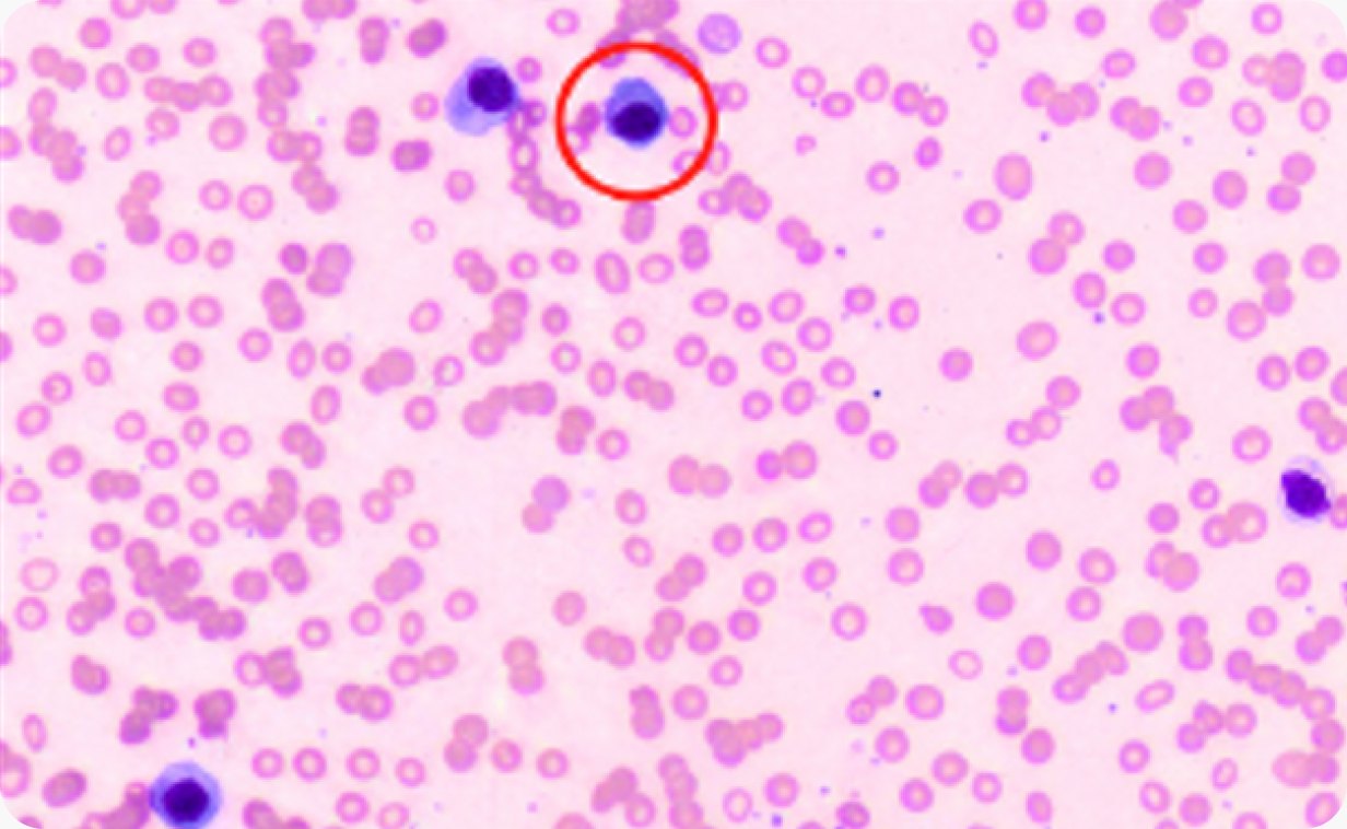
Full-Field image of PLC

Single-cell view of PLC
Challenging mantle cell lymphoma (MCL) diagnosis
A patient with hyperleukocytosis presented cells that could have been blasts or a mature lymphoid neoplasm. The AI flagged them as a mix of blasts and atypical lymphocytes. The reviewing expert reclassified, annotated, and recommended immunophenotyping by FACS analysis - all within the platform. The recommendation, annotation, and sign-off were pushed directly to the LIS.

Full-Fiel images of MCL: Her is an example of a more tricky situation. This patient has hyperleukocytosis, so a very high white blood count. You can see here on the Full-Field image that there are cells that do not belong in the peripheral blood.
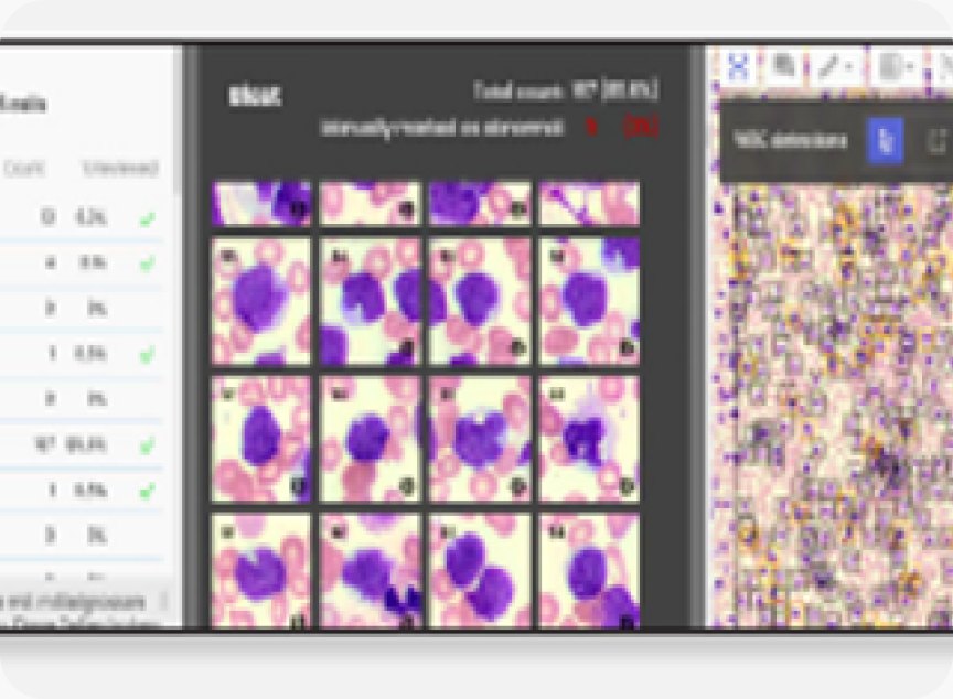
Single-cell view of MCL (blastoid variant): If we have a closer look at the differential, they look a little bit like blasts. So the AI identified some of them as blasts and some as atypical lymphocytes, so we decided to put them all together as blasts. We commented that we weren’t sure if it’s a more mature lymphoid neoplasm that looks blastoid or an acute leukemia that looks a little bit mature.

Expert’s comments from the Scopio platform to LIS: What’s important is that you can write your conclusions and recommendations. For example, the recommendation was to do Immunophenotyping by FACS analysis. You annotate and comment on the digital image and complete your report in one tool. Then, you can sign off, and everything is pushed into the LIS.
Looking ahead
ZLM is actively expanding its use of Scopio’s platform beyond peripheral blood smear review. The team has recently begun evaluating the Full-Field Bone Marrow Aspirate Application and is working with its IT department to complete LIS integration for routine use.
“In bone marrow, it’s different from the peripheral blood smear application because you are meant to count, according to WHO guidelines, at least 500 cells, which can be very time-consuming,” Richardson explains. “But with the Scopio bone marrow application, you can count thousands of cells, choose the area of scanning, and merge different scans from a single slide into the same case.”
The broader context for ZLM’s digital investment reflects a reality shared across Switzerland and the rest of the world. “The issue of lab personnel and medical staff shortages is a widespread global problem,” says Richardson. “What we are trying to achieve is to automate the laboratory process as much as we can with AI to augment the workforce and make our lives easier - not to replace it. New technology is the only way we can adapt to staff shortages while still providing quality healthcare for the patient.”
“The excellent performance of the Scopio system has completely transformed our method of blood morphology beyond our expectations - not just in our main lab, but also across all our sites. It has enabled our hematologists to work fully remotely from anywhere around the world in real time. It has accelerated diagnosis and treatment and improved the quality of our patient care.”
Dr. Lukas Graf, Head of Department of Clinical Chemistry, Hematology, Immunology Center for Laboratory Medicine (ZLM)
Related resources
More stories



- Scopio Labs’ Full-Field Peripheral Blood Smear application is CE marked and FDA-cleared, and it’s commercially available across the U.S., UK and Europe and other territories.
- Scopio’s Full-Field remote capabilities are available through the secure hospital network.