Mount Sinai Health System: Completing the Digital Hematology Workflow with Full-Field Bone Marrow Aspirate
How Mount Sinai validated and deployed Scopio’s Full-Field Bone Marrow Aspirate Application to digitize their last remaining manual hematology workflow.
Sept 2024
Validation completed
4,000+
Cases scanned since validation
11/11
Cell types validated across lineages
About Mount Sinai Health System
Mount Sinai Health System is one of the largest academic medical systems in the New York metropolitan area. It combines the Icahn School of Medicine at Mount Sinai with seven hospital campuses and more than 400 outpatient practice locations. The system has more than 9,000 physicians and more than 2,700 residents and clinical fellows, and it cares for patients from birth through geriatrics, including highly complex cases.
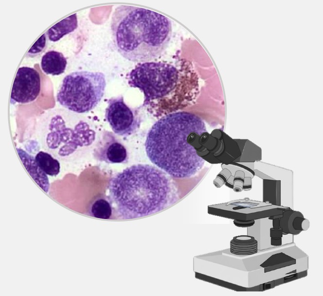
Mount Sinai’s Department of Pathology, Molecular and Cell-Based Medicine has treated digital pathology as a long-term strategic priority. In 2019, the health system announced a Digital and AI-Enabled Pathology Center of Excellence and a system-wide rollout of the Philips IntelliSite Pathology Solution. Histology and core biopsies moved to digital review. Bone marrow aspirate smears need 100x oil-immersion resolution to assess, so they stayed on the microscope and became one of the last manual steps in the hematology service.
The challenge
Mount Sinai’s Department of Pathology had already moved histology and core biopsies to digital review. Bone marrow aspirate smears were the exception. They had become one of the last manual bottlenecks in the hematology service.
A labor-intensive gold standard
Manual microscopic differential counts remain the diagnostic gold standard for bone marrow aspirates. They are also time-consuming, labor-intensive and subject to inter-observer variability. In large laboratories like Mount Sinai’s, the first pass of cell identification and differential counting falls to highly trained medical laboratory technologists (MLTs).
A shrinking technologist workforce
The national shortage of licensed MLTs, documented in the ASCP’s 2024 vacancy survey of US medical laboratories, has put growing pressure on hematology labs to do more with the same teams. The result is demand for efficiency and standardization, and for technology that can take on part of the counting workload.
“There has been an ongoing shortage of MLTs across the nation ever since the pandemic, so this calls for the adoption of new technology to augment the process of differential counting through automation.”
Overview
InstitutionMount Sinai Health System
LocationNew York, NY, USA
TypeAcademic medical system (Icahn School of Medicine at Mount Sinai)
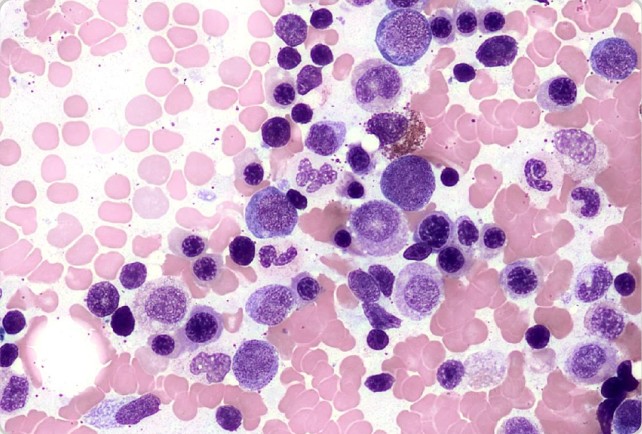
Every aspirate smear is scanned in full at 100x oil-immersion equivalent resolution. Reviewers aren’t limited to a gallery of pre-selected cells. They can move around the whole smear, assess spicules and cellularity, and zoom in on individual cells to judge cytoplasmic granularity, nuclear detail and dysplastic features, all on screen.
AI pre-classification by lineage
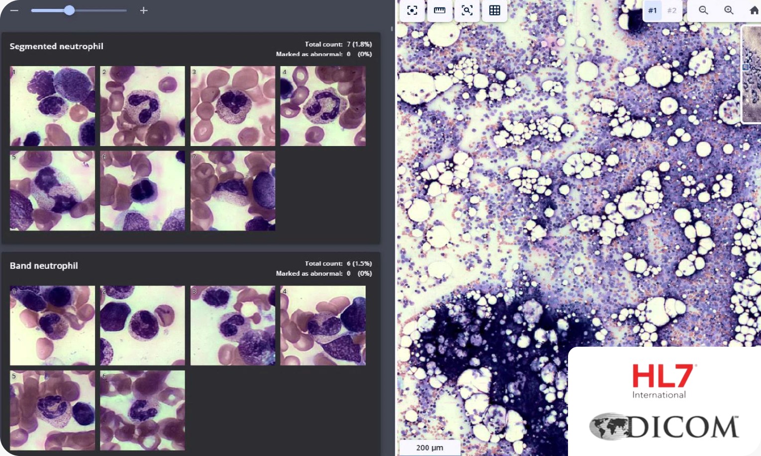
The platform’s AI-powered decision support detects cells and pre-classifies them into multiple classes. It displays them by lineage in a web-based review interface. The expert stays in control: every cell can be viewed, reclassified and annotated before sign-out, and any flagged cell can be seen in the context of the full smear.
Built into the existing digital workflow
The review interface is available through the secure hospital network. Results go to the laboratory information system via DICOM export. Mount Sinai already ran the Philips IntelliSite system for histology, so adding Scopio closed the last remaining gap without creating a separate, parallel workflow.
Cells displayed by lineage – may be viewed, reclassified and annotated
“Scopio offers a powerful ancillary tool to enhance hematopathology workflow.”
The addition of Scopio to our already existing Philips IntelliSite system marks the complete digitization of our hematology service, where bone marrow aspirate smears have historically been a bottleneck.
Validation
The team conducted a rigorous validation study with 60 bone marrow aspirate cases - 30 normal and 30 with varied morphologies reflecting their patient population. Three smears per case were stained with Wright-Giemsa; a licensed medical laboratory technologist quality-checked each set for cellularity, spicularity, and stain quality before selecting one slide for inclusion.
Accuracy was evaluated using the Rümke binomial method, comparing 200-cell AI-generated differential counts against manual reference counts performed by a licensed technologist across 11 cell types. The system passed accuracy thresholds for every cell type across the full cohort, including cases with myelodysplastic syndrome and acute myeloid leukemia.
11/11 cell types passed
200-cell differential count
Arm 1: Scopio AI-based differential count
Arm 2: Manual differential count by MLT
Supporting peer-reviewed research: Bagg A, Raess PW, Rund D, et al. Performance evaluation of a novel artificial intelligence-assisted digital microscopy system for the routine analysis of bone marrow aspirates. Modern Pathology. 2024;37(9):100542.
Clinical Impact
Since completing validation in September 2024, Mount Sinai has routinely scanned every bone marrow aspirate case, reaching over 4,000 cases. The team reports being able to appreciate cytomorphologic detail at full 100x resolution, including:
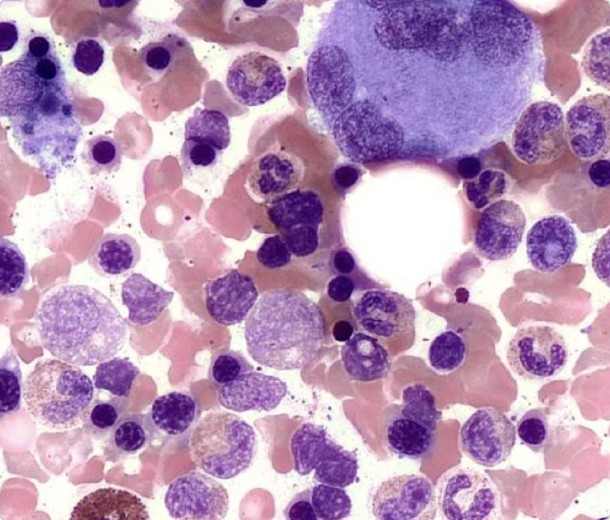
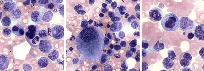
Trilineage hematopoiesis and megakaryocyte morphology
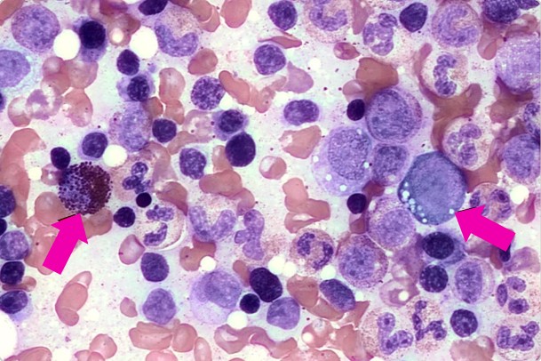
Cytoplasmic granularity and vacuolation
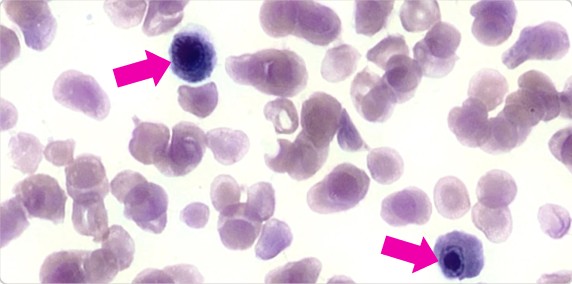
Dysplastic features including nuclear irregularity
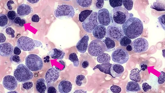
Intracytoplasmic Auer rods in leukemic promyelocytes
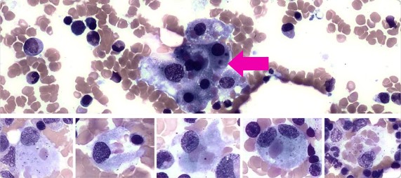
Hemophagocytosis across multiple fields
Variations in nuclear and chromatin quality
“With Full-Field bone marrow aspirate, we're able to appreciate cytoplasmic granularity, vacuolation, as well as dysplastic features.”
Trilineage hematopoiesis with megakaryocyte
Acute promyelocytic leukemia with Auer rods
Cytoplasmic granularity and vacuolation
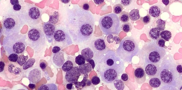
Plasma cell neoplasm
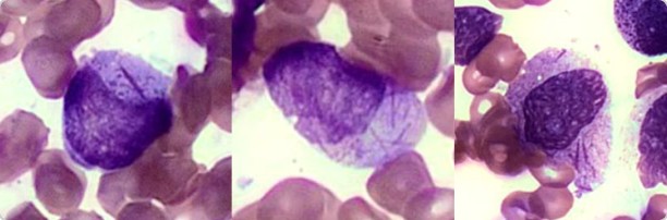
Nuclear irregularity and binucleation within the erythroid lineage
Nuclear irregularity and binucleation within the erythroid lineage
Nuclear chromatin variations
Hemophagocytosis
What’s next
Mount Sinai plans to expand their use of the platform to include Prussian blue stained slides for hemosiderin and ring sideroblast detection, as well as other sample types such as body fluids. The team is also leveraging the digital archive for technologist training, resident education, and tumor board presentations - with remote sign-out and consensus review as collaborative applications.
Scopio’s Full-Field remote capabilities are available through the secure hospital network.
Scopio’s Full-Field Bone Marrow Aspirate Application is CE-marked and cleared for sale in CE countries as well as in additional regions. Pending FDA clearance in the US.