The review of peripheral blood smear (PBS) remains a vital diagnostic tool in hematology, providing valuable insights into the morphology of blood cells and supporting diagnosis of primary and secondary blood disorders. Interpreting a PBS requires an experienced, highly trained eye; an adept understanding of blood cell morphology; and context — all of which make human expertise essential to the diagnostic process.
But technology is increasingly empowering humans to do more — to sharpen the vision of the trained eye, accelerate understanding, and expand visual context. Emerging technologies, innovative digital imaging, and AI in hematology will help scientists and hematology experts make treatment decisions faster and more confidently in the lab. That support is essential given workloads are growing faster than the pool of experts.
In a recent webinar: “Making Science Out of Art, Digitization, and Context in Full Field Morphological Hematology”, one of the world’s leading experts in hematology provided her perspective on the integration of digital blood sample review and expertise, presenting three fascinating case studies that illustrate how the art and science of diagnostic hematology can work together for the good of patients.
Wendy Erber, MD, DPhil, FRCPA, FRCPath, is a professor of pathology and laboratory medicine at the University of Western Australia and a diagnostic hematologist. Throughout her professional career, her interests have focused on adoption of new technologies, translating discoveries to improved diagnostic hematology practice, all leading to better management for patient care, especially in the field of hematological malignancies. She serves as the current chair of the International Council for Standardization in Haematology and has more than 200 publications in peer-reviewed journals, three books, and 20 book chapters to her name.
Here are a few highlights from the webinar, available on-demand here.
Morphologic hematology is an intricate art form, involving the morphological review of cells at a microscopic level and determining whether they look normal and, if not, in what way they are abnormal. That requires training, skill, and experience.
But there is subjectivity in this analysis, and inter-observer variability can lead to differences of opinion and diagnosis. In addition, it’s essential for cells to be considered in the context of their environment by looking at the cell and their surrounding areas in the blood film.
According to Dr. Erber, the future of blood film review must move from a purely manual, subjective interpretation to more detailed and standardized review, which is now possible with advanced digital morphology tools that include the full-field image scan and by leveraging advanced AI-powered decision support systems. Digital platforms also enable data and slide sharing between experts through the secure hospital network, for both diagnostic and research purposes, creating a more streamlined workflow and helping laboratories manage workloads and throughput.
In the webinar, Dr. Erber presents three cases that illustrate how expertise and technology work together to optimize the diagnostic process and decision making.
In the first case, Dr. Erber presents a 33-year-old who is 32 weeks pregnant with twins. A routine blood count showed anemia and thrombocytopenia. The initial review reported schistocytes to be present. Based on this, the provisional diagnosis was preeclampsia and she was referred for an emergency C-section at the hospital where Dr. Erber practices. . But prior to surgery, a Complete Blood Count analysis was performed including a review of her blood film that told a different story.
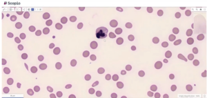
Using Scopio’s Full-Field PBS Application, Dr. Erber was able to magnify and zoom in and out on all parts of the blood smear, including the feathered edge. This comprehensive review, revealed not only some fragmented red cells, but also oval macrocytes, megaloblasts and hypersegmented neutrophils indicating megaloblastic anemia.

This diagnosis was confirmed as the lady was shown to be folate deficient. The diagnosis was revised and she was treated with folate supplementation. She did not require emergency C-section and the babies were able to remain in utero until 38 weeks.
“This total review of the blood film, being systematic in going from low to high power and reviewing a large amount of the blood film through full-field review, really changed the diagnosis,” said Dr. Erber.
In this case, full-field analysis provided a more comprehensive review of the sample and made the rare megaloblasts that had been missed in the glass slide more readily identifiable.
In another case, a 17-year-old female presents with fever, hypotension, and anemia with thrombocytopenia.

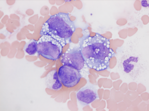
In the blood smear, key abnormal cells were detectable, but only in areas of the film that scientists typically don’t review — the edge of the film and in the thicker area of the smear where red cells are stacked. In this case, that’s where the diagnostic cells were “hiding” and why they were originally missed by the scientist looking at the glass slide through a microscope.
Fortunately, a comprehensive full-field digital review, with high magnification (100x)allowed the detection of these malignant cells, and brought the essential diagnostic indicators to light.
“These cells were escaping attention with a standard review of the blood film because we teach our scientists to stay in the monolayer and not look at the edge. And that’s where the cells were,” says Dr. Erber. “This shows the value of a full-field scan in looking for rare events that are difficult to find. If you only look in the standard part of a blood film, large cells, large events, and clumped platelets, for example, could be in that feathered end or down the side of the film. And here this was a critical diagnosis, a very serious diagnosis.” In this patient, the blood film appearances favored leukemic spread of a large cell lymphoma. This was confirmed on a follow up bone marrow examination.
Watch the webinar to learn the diagnosis in this case, see the scanned images, and get full clinical details of these cases and a third intriguing case presented by Dr. Erber.

“The accuracy of morphological analysis is really critical to reach a diagnosis and in helping our clinician colleagues treat the patient. And the decline in the number of skilled morphologists sadly presents a challenge to all of us,” concludes Dr. Erber. “But emerging technologies should help mitigate these shortages in expertise. It will enhance efficiency and standardized diagnostics through digital analysis, and patients will be the beneficiary.”