
How the CBM® Analyzer Cut Manual Morphology Review by 86% at TriCore Reference Laboratories
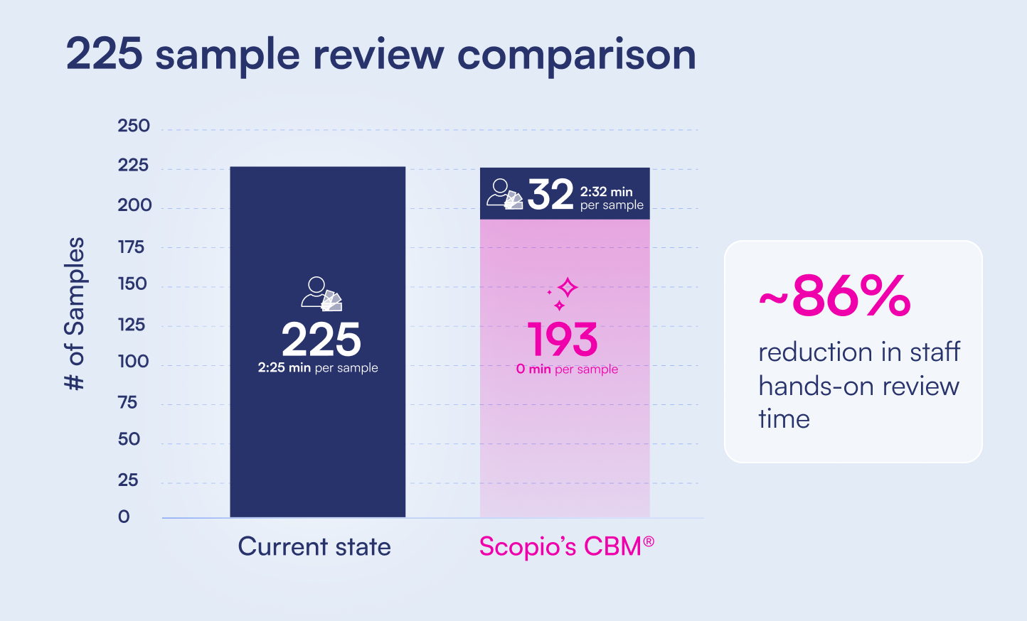
An independent time-and-motion study ran the same 225 peripheral blood smears through two workflows: TriCore's current process, and the CBM® Analyzer. Presented at ADLM 2026, alongside multi-center clinical concordance data.
What Is the CBM® Analyzer?
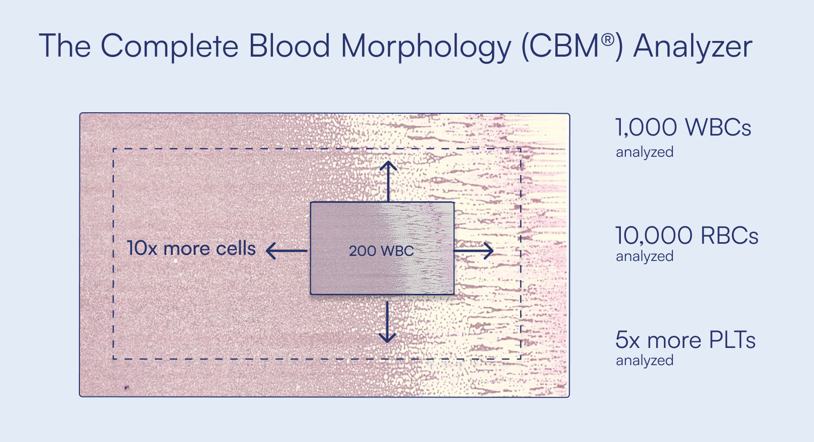
The Complete Blood Morphology (CBM®) Analyzer from Scopio Labs is the first fully autonomous analyzer for routine blood morphology reporting. It sits downstream of the CBC analyzer and eliminates the need for routine manual morphology review of peripheral blood smears. The CBM® Analyzer combines Scopio's proprietary Full-Field™ imaging with AI to quantify over 1,000 white blood cells, 10,000 red blood cells, and platelets from 50 fields of view per sample, compared with the 100 to 200 cells counted in a conventional manual differential. Samples that meet criteria are auto-reported directly to the LIS or middleware with no human review. A configurable flagging mechanism routes the remainder, including blasts, schistocytes, plasma cells and other findings each laboratory defines in its own protocols, to trained staff. Those samples are reviewed on screen using Full-Field imaging rather than at a microscope.
What Was Measured in the TriCore Workflow Comparison Study?
TriCore Reference Laboratories conducted a time-and-motion workflow comparison between its current peripheral blood smear process and the CBM® Analyzer, at its Woodward reference laboratory in Albuquerque, New Mexico. Data was collected in January and May 2026. TriCore performs 13 million billable tests per year across 16 hospital laboratories and 70+ sites, and was the first multi-site laboratory network in the nation to earn CAP 15189℠ accreditation. Its COO framed the motivation plainly.
"With laboratories nationwide facing major reductions in reimbursement, we must find ways to operate more efficiently without compromising patient care. CBM® is a timely answer to that exact challenge."
The current-state workflow
TriCore's current hematology workflow uses a Sysmex XN-9100 line with a DI-60 (CellaVision), plus manual microscopy for slides its pathology protocols will not permit on the DI-60. Of 275 slides observed, 141 (51%) required manual microscopy and 134 (49%) went to the DI-60.
"About half of our slides we use CellaVision. The other half do not meet criteria for CellaVision, so we're actually looking at those slides under the scope. When we did the future state with CBM, everything was digital."
The Woodward laboratory processes approximately 1,490 CBCs and 104 peripheral blood smears per day, at a ~7% slide review rate, running 24/7 with 1 to 2 FTEs on the review bench.
The comparison design
225 slides completed full protocol analysis in the current-state study and were then run through the CBM® workflow. The same 225 slides were used in both workflows, so no difference in case complexity could affect the comparison. Four TriCore medical technologists with no prior Scopio experience operated the analyzer, having had less than two weeks with the system. The analyzer's cell count is configurable, and TriCore ran it at its full 1,000-cell white blood cell differential rather than reducing it.
"We did the 1,000-cell differential. We didn't cheat it. You can change some of the settings, but we used the full setting."
Independent third-party analysis
All observation and timing was carried out by Nexus, an independent laboratory consulting firm engaged to conduct the time-and-motion study. Neither Scopio nor TriCore recorded the measurements. Nexus positioned observers at the DI-60 platform and the review bench simultaneously, timestamping every sample. That included system disruptions, employee breaks, shift changes, competing priorities and slide rework.
"We had a third-party organization, Nexus, come in and do all of our time studies and workflow studies. So this isn't Scopio saying this is the timing. This isn't TriCore saying it's the timing. It's a third-party group that did the timing for all of us."
Workflow Comparison Study Results
All figures below are for the 225 peripheral blood smears included in the comparison.
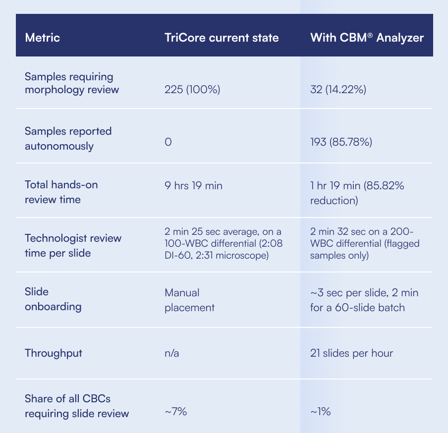
"This is the slide that really sells it for me. Of our 225 slides that we had to do manual diffs on using our old method, putting them on the CBM, we're doing 32 slides for our techs. It's about an 86% improvement overall."
Three workflow scenarios were tested: high-capacity batch runs, a 5-hour load window, and a 2-hour load window matching current state. The CBM® Analyzer flagged the same samples in every scenario and held consistent throughput regardless of batch size, loading pattern or operator.
"Essentially 21 slides an hour, and 86% of them are going to be released automatically through your systems. You can just let them go."
How much hands-on time does loading the analyzer take?
Roughly 3 seconds per slide. Slides go into 10-position cassettes, and a full 60-slide batch takes about 2 minutes to load, after which the operator walks away.
"With a full batch of 60, your tech spends 2 minutes loading it and can walk away. That probably shouldn't be a tech. That should be an assistant."
Do flagged samples take longer to review digitally?
No. The 32 flagged slides were the difficult ones by definition, samples that had already passed the Sysmex and then tripped the CBM® Analyzer's clinical flagging criteria. Technologist review averaged 2 minutes 32 seconds on screen, on par with TriCore's current manual bench at 2 minutes 31 seconds, on a harder case mix and with no walk to a scope station. Worth noting that the on-screen review used a 200-WBC differential, double the 100-WBC differential TriCore counts in its current state. This is a separate figure from the analyzer's own autonomous analysis, which counts over 1,000 white blood cells per sample.
"Essentially your techs can be just as efficient with much more complex cases. You don't actually have to take it to a scope."
Digital review is possible at that speed because Full-Field™ imaging captures the entire clinically relevant area of the slide, from the monolayer through to the feathered edge, at 100x oil immersion. A technologist can navigate the sample the way they would at a microscope.
"The image quality is just as nice as you see on the screens here. When I go through it with my pathologists, they're always very surprised to see how good it is. You can check the feathered edge for those platelet clumps. You can check the edges of the glass where they like to hide out. Anything you can do with a glass slide."
What does an 86% reduction mean in staffing terms?
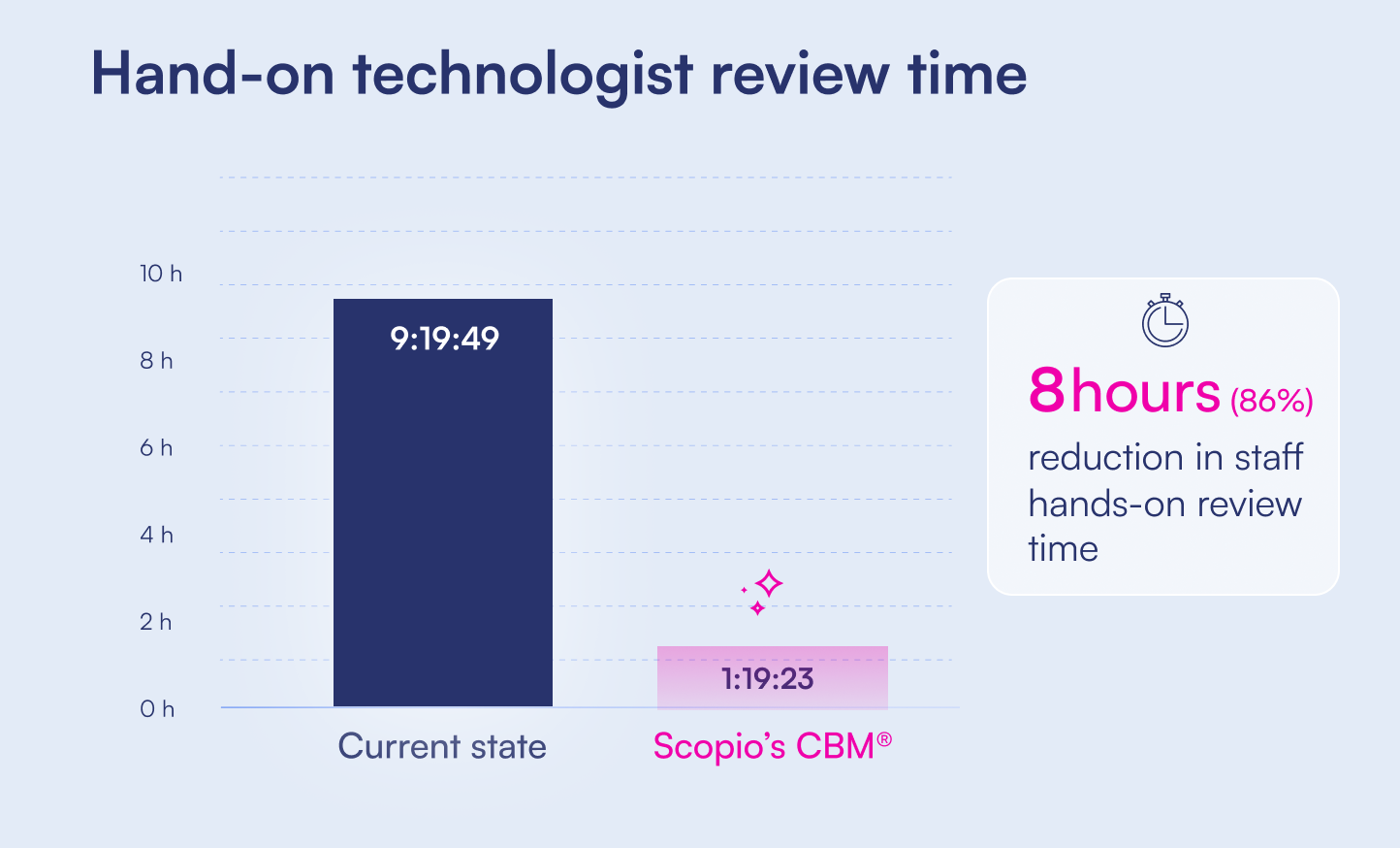
Across the 225-sample comparison, eight hours of hands-on review time were eliminated.
"We're never getting rid of FTEs. We're always short FTEs. But they're able to do something better with their time."
How Accurate Is Autonomous Morphology Analysis?
Clinical concordance data was presented from a multi-center method comparison study conducted at six laboratories: three in the US (including Brigham and Women's Hospital and UPenn), two in Europe, and one in Tel Aviv. Over 8,000 samples were processed in total across pre-submission and definitive study, with 300+ slides systematically compared against detailed manual analysis by two expert medical technologists.
Reproducibility: where manual review struggles most
Lymphocyte classification is subjective, shaped by individual interpretation of nucleus shape, cytoplasm and granules. Two expert morphologists reading the same slides quantified the problem.
"Even my morphologists with 15, 20, 30 years experience, if they both read the same slide, one's going to call something an LGL and the other will call it a regular lymph. The Pearson correlation between reviewers was 0.23."
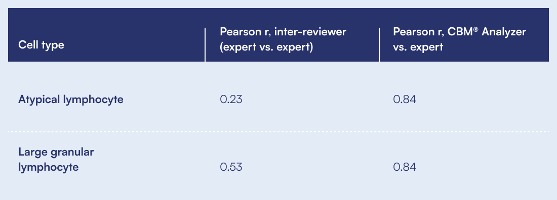
This is not a criticism of technologists. It is the nature of subjective morphological assessment at 100 to 200 cells, and it is exactly what standardization addresses.
Method comparison across cell classes
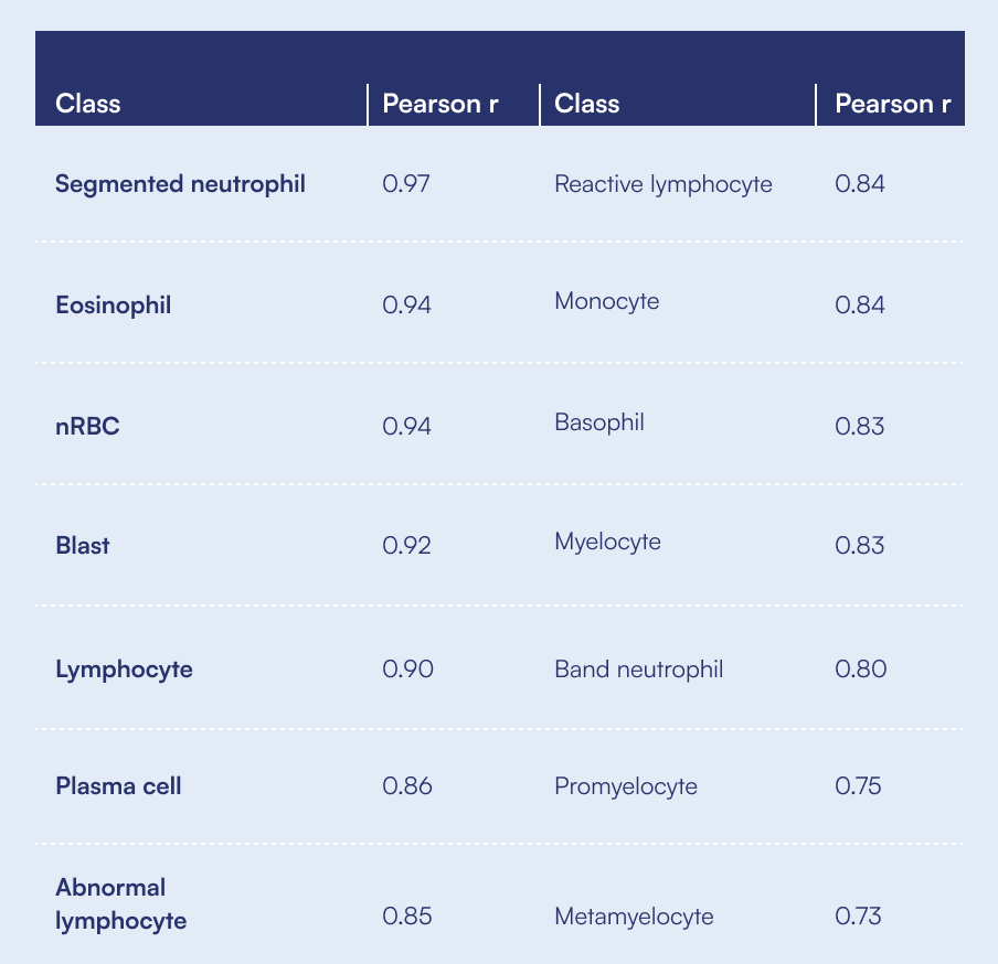
Across the full multi-center dataset, CBM® results were compared against the expert manual reference differential for each cell class.
"The blast and plasma cell correlations, 0.92 and 0.86, are just fantastic. And the ones you'd expect to be lower, your myeloid precursors and lymphocyte subsets, are also really phenomenal compared to inter-reviewer agreement."
What a 1,000-cell differential surfaces that 200 cells miss
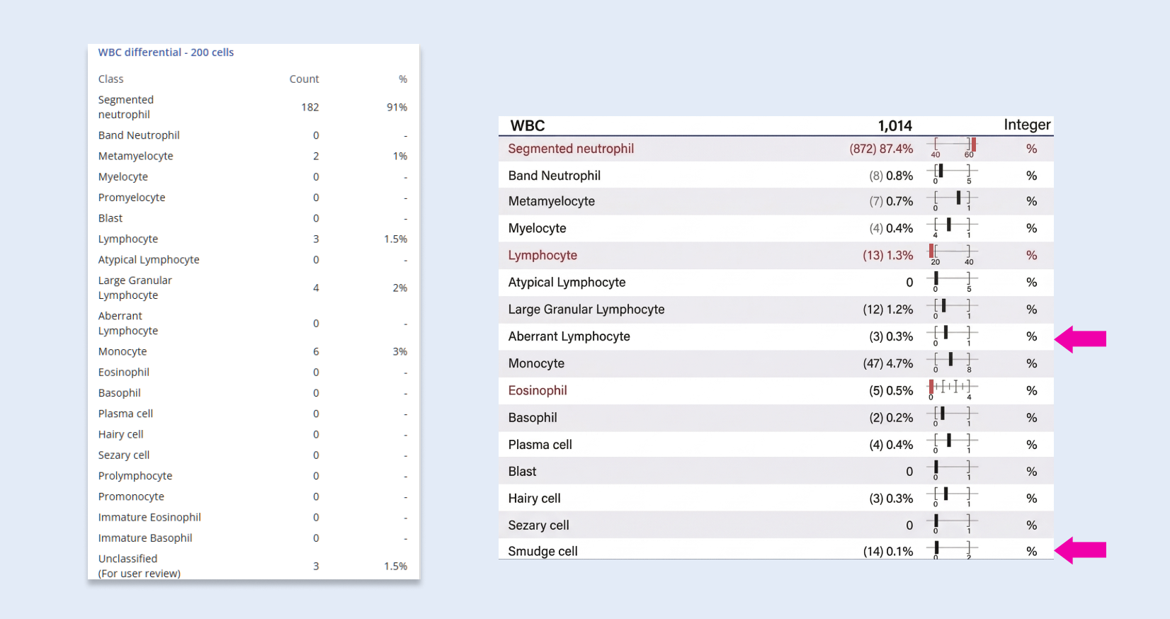
On a routine smear, results tracked closely: 75% segmented neutrophils on a 200-cell differential versus 68.6% on the CBM® Analyzer, and 12.5% lymphocytes versus 11%. On a second sample, a 200-cell differential returned 91% segmented neutrophils and 1.5% lymphocytes and nothing else of note. Counting 1,014 cells, the CBM® Analyzer surfaced a 0.3% aberrant lymphocyte population, 0.3% hairy cells, 1.2% large granular lymphocytes and smudge cells. These are sub-populations that do not reliably appear at a 200-cell sample size.
"These aren't clinically significant on their own, but it may be a great way to flag the clinician early on in a patient presenting with disease, so they can get identified and treated earlier than they would if these were the only results the clinician were getting."
Why Laboratories Are Automating Morphology Review
Roughly 25,600 US medical laboratory positions open each year against about 5,000 graduates, leaving approximately 20,000 unfilled positions annually. An estimated 25 to 35% of the MLS workforce reaches retirement age within 3 to 5 years, and test volumes are growing faster than the industry's traditional 3% baseline.
"That means you're losing 30, 40 years of experience from techs who have been doing morphology that long."
Standardization is the second driver. For multi-site health systems, autonomous analysis means consistent, defensible morphology results regardless of which laboratory ran the sample or how experienced the technologist on shift happens to be.
"If you come to one hospital as a patient, you're going to get the same numbers as you would at another hospital that may not have a 30-year morphologist on it. Maybe they've got a slew of new grads who haven't seen a blast since school."
"Automation scales expert review. That's my biggest point. It's not to replace your morphologist or technologist. It's to give them tools to do their job better."
What's Next at TriCore
- Fully implement the CBM® Analyzer at the core laboratory.
- Extend to the hospital laboratories, which process 200 to 300 peripheral blood smears per day, consolidating expertise by routing flagged slides digitally to the hematopathology team at the reference lab, with no glass shipped anywhere.
- Validate in the cancer center laboratories, where differentials are more complex and advanced analyzer features would be of most benefit.
"The system has some historical capability, so it can look back and say, 'This is the same patient who had a CLL last week, they still have a CLL this week.' We want to try that in our cancer labs, because we see the same patients over and over again."
FAQs
What percentage of blood smears can the CBM® Analyzer report autonomously?
What triggers a slide to be flagged for manual review?
How was the TriCore study compared against CellaVision?
Who conducted the time-and-motion measurements?
How many cells does the CBM® Analyzer count per sample?
Does digital review of flagged samples take longer than a microscope?
How long does it take staff to learn the system?
Is the CBM® Analyzer available for clinical use?
How much does CBM® reduce the manual review rate?
How accurate is autonomous reporting on critical findings?
Session Speakers Eric Carbonneau, Chief Operating Officer, TriCore Reference Laboratories. TriCore performs 13 million billable tests per year across 16 hospital laboratories and 70+ sites in New Mexico, and was the first multi-site laboratory network in the nation to earn CAP 15189℠ accreditation. Daniel Dees, DCLS, MLS(ASCP), Medical Director and Hematology Technical Director, Mass General Brigham. His work focuses on hematology automation, digital morphology, flow cytometry and laboratory innovation.

Recorded at ADLM 2026. See what autonomous morphology analysis would look like in your laboratory. Talk to our team or explore the Complete Blood Morphology Analyzer and Full-Field™ imaging.
All TriCore workflow figures are from a workflow comparison study at TriCore Reference Laboratory conducted by Nexus. Data collected January and May 2026. Current-state counts are based on a 100-WBC differential. CBM® flagged-sample counts are based on a 200-WBC differential.
Scopio's Complete Blood Morphology Analyzer (CBM®) is still in development, not yet available for in vitro diagnostic use. Final outputs may vary based on sample variability and final product design.