
AI and Digital Morphology in Hematology: Real Experience from Cambridge University Hospitals
How does AI change everyday work in a hematology lab, and where does it still fall short?

Clinical Summaries
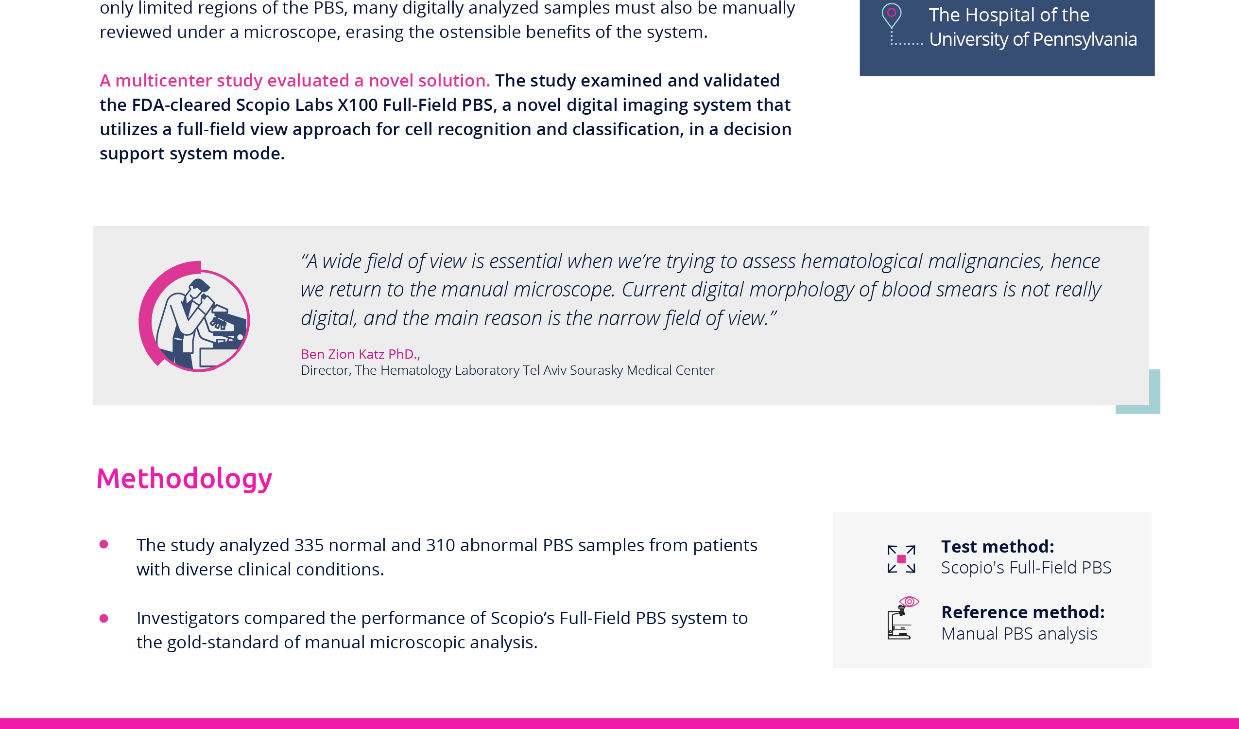
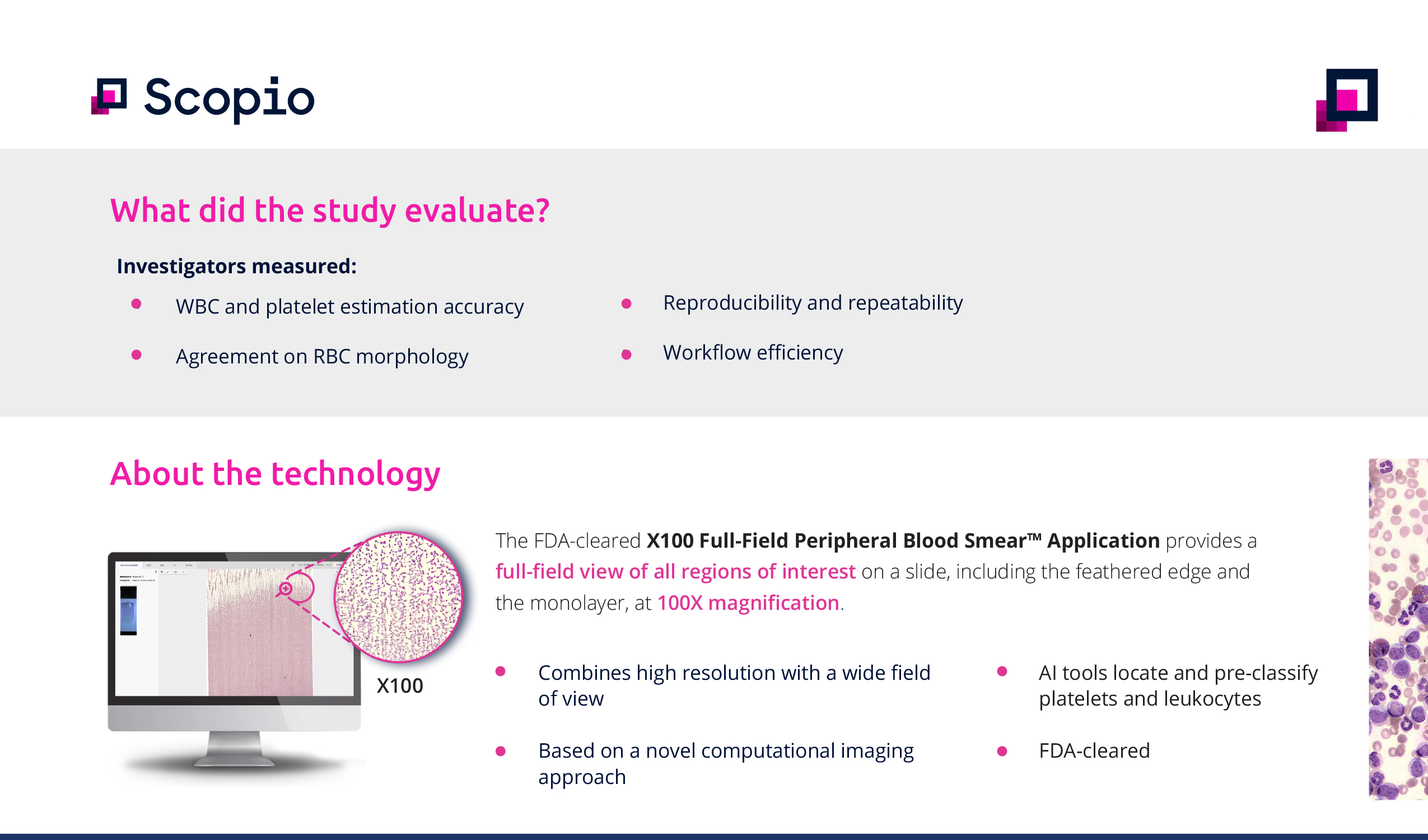
Digital cell imaging systems for peripheral blood smear analysis have improved cell and platelet recognition, but their limited field of view means many digitally analyzed samples still require manual microscopic review – erasing the efficiency gains of the system. A multicenter study evaluated Scopio’s Full-Field Peripheral Blood Smear Application, the first digital imaging system to provide a full-field view approach for cell recognition and classification, in a decision support system mode.
The study was conducted across three clinical sites: Brigham and Women’s Hospital, the Hospital of the University of Pennsylvania, and a major academic medical center.
Investigators analyzed 645 peripheral blood smear samples – 335 normal and 310 from patients with varied morphologies across diverse clinical conditions. They compared the performance of Scopio’s Full-Field Peripheral Blood Smear Application as the test method against manual microscopic analysis as the reference method. Deming regression analysis was used for comparisons of WBC and platelet estimates. Measurements of WBC and platelet estimation accuracy, agreement on RBC morphology evaluation, and reproducibility and repeatability of the system were all evaluated.
Scopio’s Full-Field Peripheral Blood Smear Application demonstrated a 60% improvement in workflow efficiency – with median review time of 7:46 minutes per case compared to 20:00 minutes for manual review.
WBC accuracy was evaluated with an efficiency of 96.29%, sensitivity of 87.68%, and specificity of 97.62%. The agreement between the test and reference method for RBC morphology reached 99.77%. Platelet estimation resulted in an efficiency of 94.89%, sensitivity of 90.00%, and specificity of 96.28%, with successful reproducibility and repeatability tests.
Scopio’s Full-Field Peripheral Blood Smear Application showed a high degree of correlation with manual microscopy across all tested parameters, with more than twice the throughput. The full-field view of specimens facilitates the long-expected disengagement between the digital application and the manual microscope – providing a path to digital morphology that eliminates the need for manual review, improves consistency, and increases operational efficiency in the hematology lab.
Published in the International Journal of Laboratory Hematology (IJLH), 2021.
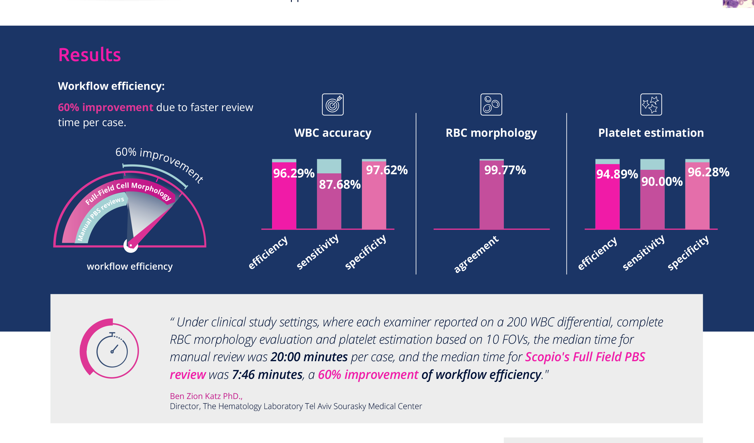
“Under clinical study settings, where each examiner reported on a 200‐WBC differential, complete RBC morphology evaluation and platelet estimation based on 10 FOVs, the median time for manual review was 20:00 minutes per case, and the median time for Scopio’s Full-Field PBS review was 7:46 minutes, a 60% improvement of workflow efficiency.”
Ben Zion Katz PhD.,
Director, The Hematology Laboratory Tel Aviv Sourasky Medical Center

Webinars
Full-Field Cell Morphology

Brochures
Full-Field Cell Morphology
Clinical Summaries
Workflow