Digital Cytology Scanner
Digital Cytology Scanner
Rapid Results and Clinical Expertise — Around-the-Clock
Faster Treatment Decisions and Better Patient Outcomes
Start experiencing more accurate test results, faster.
Reach out to your Antech Representative or fill out this short form to start experiencing Antech’s Digital Cytology service.
With our Digital Cytology service, you will run in-clinic, high-quality scans that are easily transmitted to a network of board-certified pathologists for interpretation. The scanner leverages the same best-in-class technology as the high-volume digital cytology scanners used across Antech’s North American Reference Laboratory network.
- Whole or full slide scans
- Multiple sample types
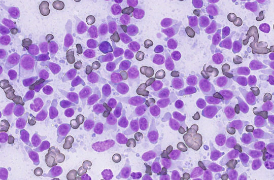
- 80x high-resolution magnification
- No slide cartridges, cover slips, immersion oil, or special staining procedures required
- Integration with 40+ Practice Management Software (PIMS) systems
- Online tutorials and user education resources
- Virtually maintenance-free
“Digital cytology has absolutely transformed our workflow and improved the client experience. I now call with official cytology results often before the client is home from their initial appointment. We’ve relied on it for immediate life threatening intra-operative case decisions. I also love the ability to get multiple pathology opinions in a matter of moments when we have a more challenging case. As we no longer rely on a mail courier for sample submission, we no longer deal with the occasional mail mis-delivery or lost sample. Our entire hospital loves the service, and I cannot see a scenario where we could go back to the old way of physical slides mailed for evaluation.”
Zachary M. Wright, DVM DACVIM (Oncology)
Chair of the VCA Pet CancerCare Alliance
Medical Director, VCA Animal Diagnostic Clinic
Transform your practice with an easy workflow that saves time and leads to faster treatment, today.
Reach out to your Antech Representative or fill out this short form to start experiencing Antech’s Digital Cytology service.
Easy to Use and Faster Access
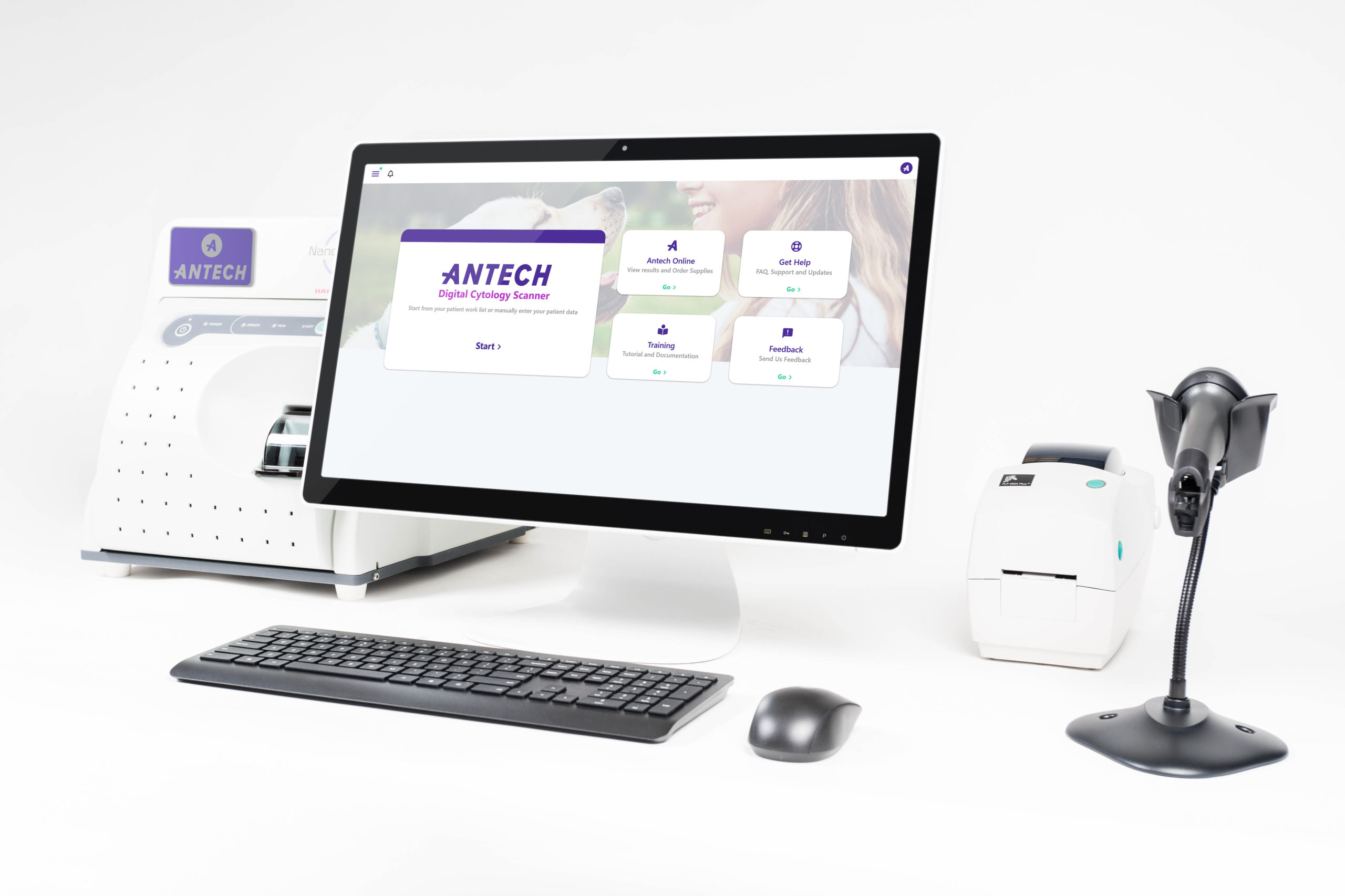
The all-in-one touchscreen display monitor, printer, barcode reader, mouse, and keyboard are included. The process is simple — quickly order, scan, and access training resources on the home screen.
24/7 Technical Service
One-click away from a Technical Support Specialist who can remotely access your system any time you need — 24/7 — or call 1-800-819-5538 (option 2) to speak to a specialist live. If you have any questions, you can access detailed instructions on the scanner’s home screen or online at: https://apps.soundvet.com/antechpoc
Real-Time Connection and Robust Results
Access to Antech’s global team of board-certified veterinary pathologists who read the slides and provided detailed reports within two hours of submission — 24 hours a day, 365 days a year. The 80x high-resolution magnification provides faster specimen identification and reduces the need for repeat scans.
No Upfront Costs
No capital equipment investment required — simply meet a low-volume monthly commitment
Integrated Solutions
Visit Antech Online to create orders, view pathology reports, and access information downloaded from your PIMS.
Acceptable Sample Types
- Fine needle aspirates from skin, subcutaneous tissue, lymph node, or viscera
- Body cavity fluid
- Fluid from cystic lesions
- Blood smear
- Urine sediment
- Bone marrow cytology for evaluation of metastasis
- Bronchoalveolar lavage/transtracheal wash fluid
- Cerebrospinal fluid
System Placement Requirements
Digital Cytology Scanner – 18.5”h x 22.5”w x 28″d, weighing ~25 lbs.
All-In-One Display Monitor – 16”h x 13.7”w x 6”d, weighing ~17 lbs.
It’s like a virtual, board-certified pathologist — right in your practice.
Watch the video to discover the impact of Antech’s Digital Cytology Service.

New customers & general inquiries
Interested in becoming an Antech customer or have a question about Antech? Please complete the form and we’ll get back to you within 24 hours.