Counting and classifying blood cells is one of the oldest laboratory methods available for the evaluation of a patients’ health. Revealing both quantitative and qualitative data, the complete blood count (CBC) and complete blood count with differential (CBC w/diff) tests are the most commonly performed tests in the clinical hematology laboratory to this day.
Counting and classifying blood cells is one of the oldest laboratory methods available for the evaluation of a patients’ health. Revealing both quantitative and qualitative data, the complete blood count (CBC) and complete blood count with differential (CBC w/diff) tests are the most commonly performed tests in the clinical hematology laboratory to this day.1
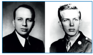
Historically, blood counting was a manual and tedious procedure. For each specimen, a technician spent on average 30 minutes at a microscope counting the red cells in a standard chamber, the result being only rarely repeatable. The need for automated hematology cell counters increased due to the burgeoning workload of laboratories seen in the 1900s.2 In 1949, Wallace H. Coulter discovered a method for detecting and counting particles suspended in a fluid medium. Forcing a particle-filled fluid through a small aperture in an electrical current modulated the current, allowing Coulter to count the number of particles in the fluid. Introduced in the mid-1950s, the Coulter Principle became the foundation of an industry responding to the need for automated hematology instruments. In 1958, Wallace and Joseph Coulter established Coulter Electronics, Inc. to pursue commercial applications of their Coulter Counter (fig 1).2
Figure 1: Coulter Brothers. Wallace H. Coulter on the left, Joseph R. Coulter on the right
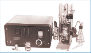
Moving from Model A (fig 2), The Model S Coulter Counter made its debut in 1969 at St Mary’s General Hospital, Portsmouth, U.K. The machine quickly made its way into the main hematology department when the results were found to be trustworthy. It was regarded as the founder of modern-day laboratory automation.2,3 Following their innovation, a revolution in hematology was underway – soon, the Coulter Counter became a staple in every hematology laboratory.
Figure 2: The Model A Coulter Counter® with the industrial version of the sample stand
By the 1980s, the field of laboratory hematology diagnostics of blood samples had advanced significantly. Flow‐based cell counters were capable of sub-classifying leukocytes and detecting abnormalities in white blood cells, red blood cells and platelets using instrument flags.4 Although the evolution in hematology workflows has continued with automated instruments increasingly performing cellular analysis and becoming an integral part of laboratory hematology diagnostics today (Fig 3), the need for morphological review of blood smears still remains. This is due to the fact that certain peripheral blood morphological characteristics do not lend themselves to evaluation by automated analyzers.4,5
Manual morphological microscopy review has remained relatively unchanged for more than a century
While the adoption of automated machines used in hematology for cell counting has become widespread, the evaluation of peripheral blood smears has challenged healthcare professionals since the invention of the microscope.5 The diagnostic relevance of cell morphology is significant, and yet the process has remained unchanged, despite advances in automated hematology.6
The examination of blood smear morphology remains indispensable to hematology, as a frontline diagnostic test to unravel the mysteries of symptoms and signs in primary and secondary hemopathies.6 One technological limitation that has remained constant, even with the latest generation of flow cytometers, is that a single cell must still pass through an aperture for analysis. To maintain laminar flow, the cell must also be sphered. As a result, automated instruments cannot easily analyze certain characteristics of cell morphology. The wide morphologic variability of many circulating hematologic cells makes it difficult for automated systems to precisely characterize them . Most analyzers, however, can assist in characterizing these cells by pre-classifying them as abnormal (through large unstained cell classification or flagging) and prompting a manual slide review.5
Morphological analysis of the Romanowsky‐stained peripheral blood smear is an essential element in the diagnosis of hematological diseases and conditions.4 Traditionally, morphological analysis of the blood smear has been performed by manual light microscopy, representing one of the last bastions of analog technology yet to undergo digital transformation. The process of viewing, annotating, comparing and analyzing specimens on glass slides has, until recently, remained relatively unchanged for more than a century.7 Although this method is the gold standard, it has the disadvantages of being labor‐intensive, requiring continuous training of personnel, and being subject to relatively large interobserver variability. It is difficult to access secondary consultations using the classical manual microscopy method and storage of glass slides remains a challenge.4 Thus, there is an increased demand for the development of high-resolution digital microscopy systems capable of performing morphological analysis of blood smears in an automated manner.4
Looking ahead: the digital cell morphology revolution
The advantages of automation in hematology are clearly seen through the innovations of the Coulter brothers. Taking cell counting from a manual and labor-intensive process to one that is done quickly, efficiently and reliably, changed the standard for hematology laboratories the world over. The industry is anxiously awaiting the second-wave revolution in hematology automation, bringing cell morphology into the digital age.
Seven solutions: How the most innovative hematology labs are tackling their top threats.
Hematology laboratories around the world face similar operational and organizational challenges. A shrinking workforce, increasing testing volumes and case complexity, and manual processes are among the virtually universal barriers to efficiency and quality service.
Nearly a quarter of all hematology labs analyze more than 100 peripheral blood smears (PBS) every day, yet the vast majority are still performed manually using an optical microscope. That disturbing statistic is just one creating a growing crisis in the hematology lab. In the age of widespread digitization in healthcare and across industries, hematology laboratory procedures are falling behind. And it's that misalignment that planted the seeds of Scopio Labs.
Something like 70% of today’s medical decisions depends on laboratory test results, and clinical laboratories therefore play an absolutely essential role in today’s healthcare system. Yet the most common hematology lab tests like the peripheral blood smear review are still mainly conducted manually with a microscope. They require classifying and counting cells by type and hunting for abnormalities. Does it work? Sure. Is it the best we can do? Not by a long shot. This method is labor intensive, time-consuming, requires continuous staff training, and is subject to relatively large inter-observer variability